1SFU

Crystal structure of the viral Zalpha domain bound to left-handed Z-DNA


Experimental Data Snapshot

  • Method: X-RAY DIFFRACTION
  • Resolution: 2.00 Å
  • R-Value Free: 0.270 
  • R-Value Work: 0.237 

wwPDB Validation   3D Report Full Report


This is version 1.3 of the entry. See complete history


Literature

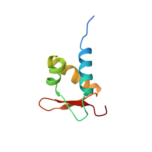
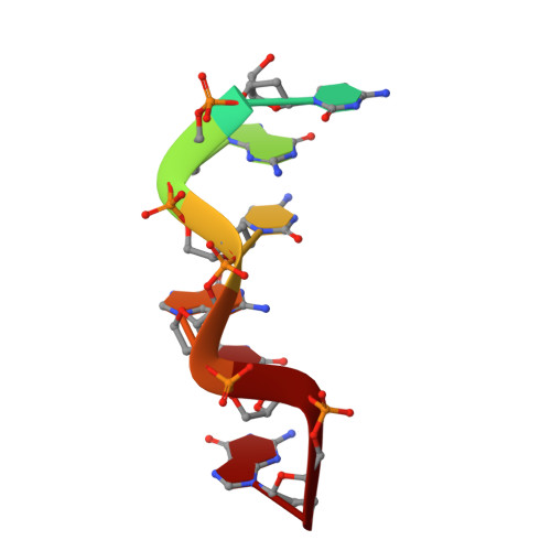
A poxvirus protein forms a complex with left-handed Z-DNA: crystal structure of a Yatapoxvirus Zalpha bound to DNA.

Ha, S.C.Lokanath, N.K.Van Quyen, D.Wu, C.A.Lowenhaupt, K.Rich, A.Kim, Y.G.Kim, K.K.

(2004) Proc Natl Acad Sci U S A 101: 14367-14372

  • DOI: https://doi.org/10.1073/pnas.0405586101
  • Primary Citation of Related Structures:  
    1SFU

  • PubMed Abstract: 

    A conserved feature of poxviruses is a protein, well characterized as E3L in vaccinia virus, that confers IFN resistance on the virus. This protein comprises two domains, an N-terminal Z-DNA-binding protein domain (Zalpha) and a C-terminal double-stranded RNA-binding domain. Both are required for pathogenicity of vaccinia virus in mice infected by intracranial injection. Here, we describe the crystal structure of the Zalpha domain from the E3L-like protein of Yaba-like disease virus, a Yatapoxvirus, in a complex with Z-DNA, solved at a 2.0-A resolution. The DNA contacting surface of Yaba-like disease virus Zalpha(E3L) closely resembles that of other structurally defined members of the Zalpha family, although some variability exists in the beta-hairpin region. In contrast to the Z-DNA-contacting surface, the nonbinding surface of members of the Zalpha family are unrelated; this surface may effect protein-specific interactions. The presence of the conserved and tailored Z-DNA-binding surface, which interacts specifically with the zigzag backbone and syn base diagnostic of the Z-form, reinforces the importance to poxvirus infection of the ability of this protein to recognize the Z-conformation.


  • Organizational Affiliation

    Department of Molecular Cell Biology, Samsung Biomedical Research Institute, Sungkyunkwan University School of Medicine, Suwon 440-746, Korea.


Macromolecules

Find similar proteins by:  (by identity cutoff)  |  3D Structure
Entity ID: 2
MoleculeChains Sequence LengthOrganismDetailsImage
34L proteinC [auth A],
D [auth B]
75Yaba-like disease virusMutation(s): 0 
UniProt
Find proteins for Q9DHS8 (Yaba-like disease virus)
Explore Q9DHS8 
Go to UniProtKB:  Q9DHS8
Entity Groups  
Sequence Clusters30% Identity50% Identity70% Identity90% Identity95% Identity100% Identity
UniProt GroupQ9DHS8
Sequence Annotations
Expand
  • Reference Sequence

Find similar nucleic acids by:  Sequence   |   3D Structure  

Entity ID: 1
MoleculeChains LengthOrganismImage
5'-D(*T*CP*GP*CP*GP*CP*G)-3'A [auth C],
B [auth D]
7N/A
Sequence Annotations
Expand
  • Reference Sequence
Experimental Data & Validation

Experimental Data

  • Method: X-RAY DIFFRACTION
  • Resolution: 2.00 Å
  • R-Value Free: 0.270 
  • R-Value Work: 0.237 
  • Space Group: P 21 21 2
Unit Cell:
Length ( Å )Angle ( ˚ )
a = 51.203α = 90
b = 92.452β = 90
c = 48.012γ = 90
Software Package:
Software NamePurpose
HKL-2000data collection
SCALEPACKdata scaling
SOLVEphasing
CNSrefinement
HKL-2000data reduction

Structure Validation

View Full Validation Report


Entry History 

Deposition Data

Revision History  (Full details and data files)

  • Version 1.0: 2004-08-17
    Type: Initial release
  • Version 1.1: 2008-04-29
    Changes: Version format compliance
  • Version 1.2: 2011-07-13
    Changes: Version format compliance
  • Version 1.3: 2024-02-14
    Changes: Data collection, Database references