1SCU

THE CRYSTAL STRUCTURE OF SUCCINYL-COA SYNTHETASE FROM ESCHERICHIA COLI AT 2.5 ANGSTROMS RESOLUTION


Experimental Data Snapshot

  • Method: X-RAY DIFFRACTION
  • Resolution: 2.50 Å
  • R-Value Work: 0.216 
  • R-Value Observed: 0.216 

wwPDB Validation   3D Report Full Report


This is version 1.4 of the entry. See complete history


Literature

The crystal structure of succinyl-CoA synthetase from Escherichia coli at 2.5-A resolution.

Wolodko, W.T.Fraser, M.E.James, M.N.Bridger, W.A.

(1994) J Biol Chem 269: 10883-10890

  • DOI: https://doi.org/10.2210/pdb1scu/pdb
  • Primary Citation of Related Structures:  
    1SCU

  • PubMed Abstract: 

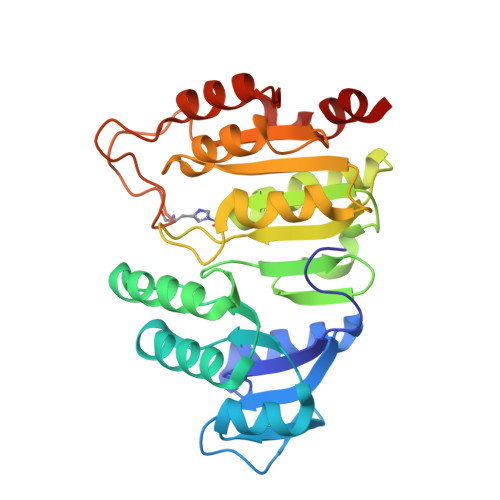
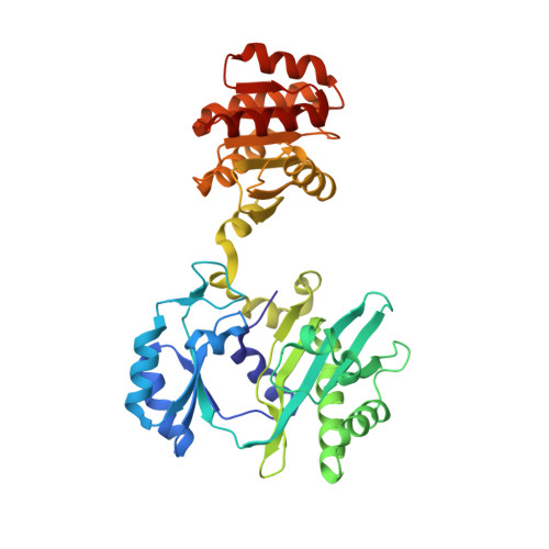
    The x-ray crystal structure of succinyl-CoA synthetase (SCS) from Escherichia coli has been determined by the method of multiple isomorphous replacement to a resolution of 2.5 A. Crystals of SCS are tetragonal with a space group of P4(3)22 and unit cell dimensions of a = b = 98.47 A and c = 400.6 A. One molecule of SCS (142 kDa) is contained in the asymmetric unit. The current model has been refined to a conventional R factor of 21.6% with root mean square deviations from ideal stereochemistry of 0.022 A for bond lengths and 3.25 degrees for bond angles. The quaternary organization of the E. coli enzyme is an alpha 2 beta 2 heterotetramer. In this tetramer, the alpha-subunits interact only with the beta-subunits, whereas the beta-subunits interact to form the dimer of alpha beta-dimers. The two active site pockets are located at regions of contact between alpha- and beta-subunits. One molecule of coenzyme A is bound to each alpha-subunit at a typical nucleotide-binding motif, and His-246 of each alpha-subunit is phosphorylated. This phosphohistidine, a catalytic intermediate, is stabilized by two helix dipoles (the "power" helices), one from each of the two subunit types. A short segment of the beta-subunit from one alpha beta-dimer is in close proximity to the CoA-binding site of the other alpha beta-dimer, providing a possible rationale for the overall tetrameric structure.


  • Organizational Affiliation

    Department of Biochemistry, University of Alberta, Edmonton, Canada.


Macromolecules
Find similar proteins by:  (by identity cutoff)  |  3D Structure
Entity ID: 1
MoleculeChains Sequence LengthOrganismDetailsImage
SUCCINYL-COA SYNTHETASE, ALPHA SUBUNITA,
C [auth D]
288Escherichia coliMutation(s): 0 
EC: 6.2.1.5
UniProt
Find proteins for P0AGE9 (Escherichia coli (strain K12))
Explore P0AGE9 
Go to UniProtKB:  P0AGE9
Entity Groups  
Sequence Clusters30% Identity50% Identity70% Identity90% Identity95% Identity100% Identity
UniProt GroupP0AGE9
Sequence Annotations
Expand
  • Reference Sequence
Find similar proteins by:  (by identity cutoff)  |  3D Structure
Entity ID: 2
MoleculeChains Sequence LengthOrganismDetailsImage
SUCCINYL-COA SYNTHETASE, BETA SUBUNITB,
D [auth E]
388Escherichia coliMutation(s): 0 
EC: 6.2.1.5
UniProt
Find proteins for P0A836 (Escherichia coli (strain K12))
Explore P0A836 
Go to UniProtKB:  P0A836
Entity Groups  
Sequence Clusters30% Identity50% Identity70% Identity90% Identity95% Identity100% Identity
UniProt GroupP0A836
Sequence Annotations
Expand
  • Reference Sequence
Small Molecules
Ligands 1 Unique
IDChains Name / Formula / InChI Key2D Diagram3D Interactions
COA
Query on COA

Download Ideal Coordinates CCD File 
E [auth A],
F [auth D]
COENZYME A
C21 H36 N7 O16 P3 S
RGJOEKWQDUBAIZ-IBOSZNHHSA-N
Modified Residues  1 Unique
IDChains TypeFormula2D DiagramParent
NEP
Query on NEP
A,
C [auth D]
L-PEPTIDE LINKINGC6 H10 N3 O5 PHIS
Experimental Data & Validation

Experimental Data

  • Method: X-RAY DIFFRACTION
  • Resolution: 2.50 Å
  • R-Value Work: 0.216 
  • R-Value Observed: 0.216 
  • Space Group: P 43 2 2
Unit Cell:
Length ( Å )Angle ( ˚ )
a = 98.47α = 90
b = 98.47β = 90
c = 400.6γ = 90
Software Package:
Software NamePurpose
X-PLORmodel building
TNTrefinement
X-PLORrefinement
X-PLORphasing

Structure Validation

View Full Validation Report


Entry History 

Deposition Data

Revision History  (Full details and data files)

  • Version 1.0: 1995-04-20
    Type: Initial release
  • Version 1.1: 2008-03-03
    Changes: Version format compliance
  • Version 1.2: 2011-07-13
    Changes: Version format compliance
  • Version 1.3: 2019-07-17
    Changes: Data collection, Derived calculations, Other, Refinement description
  • Version 1.4: 2019-08-14
    Changes: Data collection, Refinement description