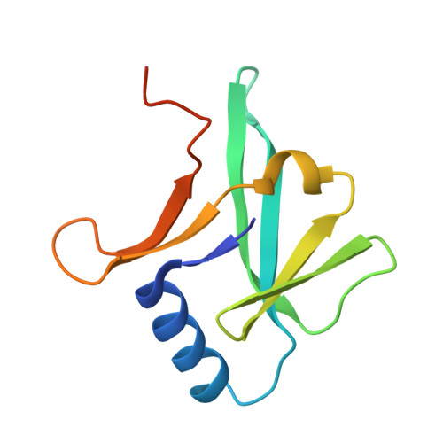
Crystal structure of IscA, an iron-sulfur cluster assembly protein from Escherichia coli.
Cupp-Vickery, J.R., Silberg, J.J., Ta, D.T., Vickery, L.E.(2004) J Mol Biol 338: 127-137
- PubMed: 15050828
- DOI: https://doi.org/10.1016/j.jmb.2004.02.027
- Primary Citation of Related Structures:
1S98 - PubMed Abstract:
IscA, an 11 kDa member of the hesB family of proteins, binds iron and [2Fe-2S] clusters, and participates in the biosynthesis of iron-sulfur proteins. We report the crystal structure of the apo-protein form of IscA from Escherichia coli to a resolution of 2.3A. The crystals belong to the space group P3(2)21 and have unit cell dimensions a=b=66.104 A, c=150.167 A (alpha=beta=90 degrees, gamma=120 degrees ). The structure was solved using single-wavelength anomalous dispersion (SAD) phasing of a selenomethionyl derivative, and the IscA model was refined to R=21.4% (Rfree=25.4%). IscA exists as an (alpha1alpha2)2 homotetramer with the (alpha1alpha2) dimer comprising the asymmetric unit. Cys35, implicated in Fe-S cluster assembly, is located in a central cavity formed at the tetramer interface with the gamma-sulfur atoms of residues from the alpha1 and alpha2' monomers (and alpha1'alpha2) positioned close to one another (approximately equal 7 A). C-terminal residues 99-107 are disordered, and the exact positions of Cys99 and Cys101 could not be determined. However, computer modeling of C-terminal residues in the tetramer suggests that Cys99 and Cys101 in the alpha1 monomer and those of the alpha1' monomer (or alpha2 and alpha2') are positioned sufficiently close to coordinate [2Fe-2S] clusters between the two dimers, whereas this is not possible within the (alpha1alpha2) or (alpha1'alpha2') dimer. This symmetrical arrangement allows for binding of two [2Fe-2S] clusters on opposite sides of the tetramer. Modeling further reveals that Cys101 is positioned sufficiently close to Cys35 to allow Cys35 to participate in cluster assembly, formation, or transfer.
Organizational Affiliation:
Department of Physiology and Biophysics, University of California, Irvine, CA 92697, USA. jvickery@uci.edu