1S80

Structure of Serine Acetyltransferase from Haemophilis influenzae Rd


Experimental Data Snapshot

  • Method: X-RAY DIFFRACTION
  • Resolution: 2.70 Å
  • R-Value Free: 0.219 
  • R-Value Work: 0.172 
  • R-Value Observed: 0.174 

wwPDB Validation   3D Report Full Report


This is version 1.3 of the entry. See complete history


Literature

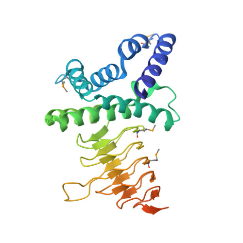
Structure of serine acetyltransferase from Haemophilus influenzae Rd.

Gorman, J.Shapiro, L.

(2004) Acta Crystallogr D Biol Crystallogr 60: 1600-1605

  • DOI: https://doi.org/10.1107/S0907444904015240
  • Primary Citation of Related Structures:  
    1S80

  • PubMed Abstract: 

    The crystal structure of serine acetyltransferase (SAT) from Haemophilus influenzae Rd determined at 2.7 A resolution is presented. SAT is a member of a family of hexapeptide-containing transferases that contain six-residue tandem repeats (LIV)-G-X(4) that have been shown to form left-handed parallel beta-helices. In the current structure, each protomer is comprised of two domains: an N-terminal alpha-helical domain and a C-terminal left-handed parallel beta-helix domain. Although other members of this protein family are known to form trimeric structures, SAT forms a dimer of trimers in which the trimer interface is mediated through interactions between both the beta-helix domains and N-terminal domains; these trimers dimerize through contacts in the N-terminal domain. All dimer-of-trimer interactions are mediated through amino acids within an N-terminal extension common only to a subset of SATs, suggesting that members of this subfamily may also adopt hexameric structures. Putative active sites are formed by crevices between adjacent protomers in a trimer. Thus, six independent active sites exist in the hexameric enzyme complex.


  • Organizational Affiliation

    Department of Ophthalmology, Columbia University College of Physicians and Surgeons, 630 West 168th Street, New York, NY 10032, USA.


Macromolecules
Find similar proteins by:  (by identity cutoff)  |  3D Structure
Entity ID: 1
MoleculeChains Sequence LengthOrganismDetailsImage
Serine acetyltransferase
A, B, C, D, E
A, B, C, D, E, F
278Haemophilus influenzaeMutation(s): 4 
Gene Names: CYSEHI0606
EC: 2.3.1.30
UniProt
Find proteins for P43886 (Haemophilus influenzae (strain ATCC 51907 / DSM 11121 / KW20 / Rd))
Explore P43886 
Go to UniProtKB:  P43886
Entity Groups  
Sequence Clusters30% Identity50% Identity70% Identity90% Identity95% Identity100% Identity
UniProt GroupP43886
Sequence Annotations
Expand
  • Reference Sequence
Small Molecules
Modified Residues  1 Unique
IDChains TypeFormula2D DiagramParent
MSE
Query on MSE
A, B, C, D, E
A, B, C, D, E, F
L-PEPTIDE LINKINGC5 H11 N O2 SeMET
Experimental Data & Validation

Experimental Data

  • Method: X-RAY DIFFRACTION
  • Resolution: 2.70 Å
  • R-Value Free: 0.219 
  • R-Value Work: 0.172 
  • R-Value Observed: 0.174 
  • Space Group: C 2 2 21
Unit Cell:
Length ( Å )Angle ( ˚ )
a = 129.963α = 90
b = 209.67β = 90
c = 136.569γ = 90
Software Package:
Software NamePurpose
REFMACrefinement
DENZOdata reduction
SCALEPACKdata scaling
SOLVEphasing

Structure Validation

View Full Validation Report


Entry History 

Deposition Data

Revision History  (Full details and data files)

  • Version 1.0: 2004-08-31
    Type: Initial release
  • Version 1.1: 2008-04-29
    Changes: Version format compliance
  • Version 1.2: 2011-07-13
    Changes: Version format compliance
  • Version 1.3: 2021-02-03
    Changes: Database references, Derived calculations, Structure summary