1S5O

Structural and Mutational Characterization of L-carnitine Binding to Human carnitine Acetyltransferase


Experimental Data Snapshot

  • Method: X-RAY DIFFRACTION
  • Resolution: 1.80 Å
  • R-Value Work: 0.189 
  • R-Value Observed: 0.189 

wwPDB Validation   3D Report Full Report


This is version 1.3 of the entry. See complete history


Literature

Structural and mutational characterization of l-carnitine binding to human carnitine acetyltransferase.

Govindasamy, L.Kukar, T.Lian, W.Pedersen, B.Gu, Y.Agbandje-McKenna, M.Jin, S.McKenna, R.Wu, D.

(2004) J Struct Biol 146: 416-424

  • DOI: https://doi.org/10.1016/j.jsb.2004.01.011
  • Primary Citation of Related Structures:  
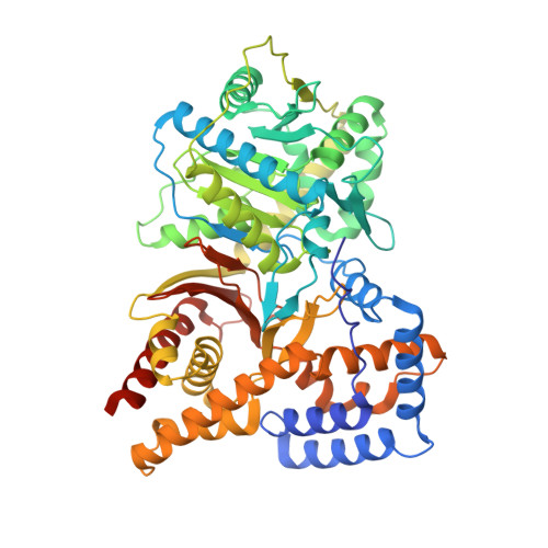
    1S5O

  • PubMed Abstract: 

    We report the crystal structure of a binary complex of human peroxisomal carnitine acetyltransferase and the substrate l-carnitine, refined to a resolution of 1.8 Angstrom with an R(factor) value of 18.9% (R(free)=22.3%). L-carnitine binds to a preformed pocket in the active site tunnel of carnitine acetyltransferase aligned with His(322). The quaternary nitrogen of carnitine forms a pi-cation interaction with Phe(545), while Arg(497) forms an electrostatic interaction with the negatively charged carboxylate group. An extensive hydrogen bond network also occurs between the carboxylate group and Tyr(431), Thr(444), and a bound water molecule. Site-directed mutagenesis and kinetic characterization reveals that Tyr(431), Thr(444), Arg(497), and Phe(545) are essential for high affinity binding of L-carnitine.


  • Organizational Affiliation

    Biochemistry and Molecular Biology, University of Florida and The McKnight Brain Institute, 1600 Archer Rd., Gainesville, FL 32610, USA.


Macromolecules
Find similar proteins by:  (by identity cutoff)  |  3D Structure
Entity ID: 1
MoleculeChains Sequence LengthOrganismDetailsImage
carnitine acetyltransferase isoform 2616Homo sapiensMutation(s): 0 
EC: 2.3.1.7
UniProt & NIH Common Fund Data Resources
Find proteins for P43155 (Homo sapiens)
Explore P43155 
Go to UniProtKB:  P43155
PHAROS:  P43155
GTEx:  ENSG00000095321 
Entity Groups  
Sequence Clusters30% Identity50% Identity70% Identity90% Identity95% Identity100% Identity
UniProt GroupP43155
Sequence Annotations
Expand
  • Reference Sequence
Small Molecules
Ligands 1 Unique
IDChains Name / Formula / InChI Key2D Diagram3D Interactions
152
Query on 152

Download Ideal Coordinates CCD File 
B [auth A]CARNITINE
C7 H16 N O3
PHIQHXFUZVPYII-ZCFIWIBFSA-O
Experimental Data & Validation

Experimental Data

  • Method: X-RAY DIFFRACTION
  • Resolution: 1.80 Å
  • R-Value Work: 0.189 
  • R-Value Observed: 0.189 
  • Space Group: P 21 21 21
Unit Cell:
Length ( Å )Angle ( ˚ )
a = 137.56α = 90
b = 84.65β = 90
c = 57.37γ = 90
Software Package:
Software NamePurpose
DENZOdata reduction
SCALEPACKdata scaling
CNSrefinement
SHELXL-97refinement
CNSphasing

Structure Validation

View Full Validation Report


Entry History 

Deposition Data

Revision History  (Full details and data files)

  • Version 1.0: 2004-02-03
    Type: Initial release
  • Version 1.1: 2008-04-29
    Changes: Version format compliance
  • Version 1.2: 2011-07-13
    Changes: Version format compliance
  • Version 1.3: 2023-08-23
    Changes: Data collection, Database references, Derived calculations, Refinement description