1S44

The structure and refinement of apocrustacyanin C2 to 1.6A resolution and the search for differences between this protein and the homologous apoproteins A1 and C1.


Experimental Data Snapshot

  • Method: X-RAY DIFFRACTION
  • Resolution: 1.60 Å
  • R-Value Free: 0.247 
  • R-Value Work: 0.203 
  • R-Value Observed: 0.205 

wwPDB Validation   3D Report Full Report


This is version 1.4 of the entry. See complete history


Literature

The structure and refinement of apocrustacyanin C2 to 1.3 A resolution and the search for differences between this protein and the homologous apoproteins A1 and C1.

Habash, J.Helliwell, J.R.Raftery, J.Cianci, M.Rizkallah, P.J.Chayen, N.E.Nneji, G.A.Zagalsky, P.F.

(2004) Acta Crystallogr D Biol Crystallogr 60: 493-498

  • DOI: https://doi.org/10.1107/S090744490400037X

  • PubMed Abstract: 

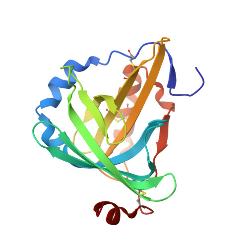
    The blue carotenoprotein alpha-crustacyanin of Homarus gammarus lobster carapace is comprised chemically of five 20 kDa subunits. Only two genes for the proteins have been isolated (J. B. C. Findlay, personal communication) and the five apoproteins fall into two sets of homologous proteins based on their chemical properties (CRTC, consisting of apoproteins C(1), C(2) and A(1), and CRTA, consisting of apoproteins A(2) and A(3)). The diffraction quality of apo C(2) has been improved from 2.2 to 1.3 A and its structure solved. The structure is compared with the A(1) and C(1) proteins determined at 1.4 A [Cianci et al. (2001), Acta Cryst. D57, 1219-1229] and 1.15 A, respectively [Gordon et al. (2001), Acta Cryst. D57, 1230-1237] and found to be very similar. Normalized B-factor difference plots per residue of different types were used to try to find chemically modified residues; none were found at these resolutions. It remains possible that the differences between the CRTC proteins result from differences in amidation. By comparison of a crystal grown with glycerol (studied at 1.6 A) and one grown without glycerol (studied at 1.3 A) it was seen that glycerol bound at the astaxanthin site.


  • Organizational Affiliation

    Section of Structural Chemistry, Department of Chemistry, University of Manchester, Manchester M13 9PL, England.


Macromolecules
Find similar proteins by:  (by identity cutoff)  |  3D Structure
Entity ID: 1
MoleculeChains Sequence LengthOrganismDetailsImage
Crustacyanin A1 subunit
A, B
180Homarus gammarusMutation(s): 0 
UniProt
Find proteins for P58989 (Homarus gammarus)
Explore P58989 
Go to UniProtKB:  P58989
Entity Groups  
Sequence Clusters30% Identity50% Identity70% Identity90% Identity95% Identity100% Identity
UniProt GroupP58989
Sequence Annotations
Expand
  • Reference Sequence
Experimental Data & Validation

Experimental Data

  • Method: X-RAY DIFFRACTION
  • Resolution: 1.60 Å
  • R-Value Free: 0.247 
  • R-Value Work: 0.203 
  • R-Value Observed: 0.205 
  • Space Group: P 21 21 21
Unit Cell:
Length ( Å )Angle ( ˚ )
a = 41.167α = 90
b = 79.879β = 90
c = 109.562γ = 90
Software Package:
Software NamePurpose
REFMACrefinement
MOSFLMdata reduction
CCP4data scaling

Structure Validation

View Full Validation Report


Entry History 

Deposition Data

Revision History  (Full details and data files)

  • Version 1.0: 2004-04-27
    Type: Initial release
  • Version 1.1: 2008-04-29
    Changes: Version format compliance
  • Version 1.2: 2011-07-13
    Changes: Non-polymer description, Version format compliance
  • Version 1.3: 2018-04-04
    Changes: Data collection
  • Version 1.4: 2023-08-23
    Changes: Data collection, Database references, Derived calculations, Refinement description