The crystal structure of the hydrolase domain of 10-formyltetrahydrofolate dehydrogenase: mechanism of hydrolysis and its interplay with the dehydrogenase domain.
Chumanevich, A.A., Krupenko, S.A., Davies, C.(2004) J Biol Chem 279: 14355-14364
- PubMed: 14729668
- DOI: https://doi.org/10.1074/jbc.M313934200
- Primary Citation of Related Structures:
1S3I - PubMed Abstract:
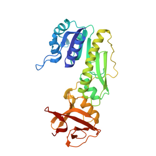
10-Formyltetrahydrofolate dehydrogenase (FDH) converts 10-formyltetrahydrofolate, a precursor for nucleotide biosynthesis, to tetrahydrofolate. The protein comprises two functional domains: a hydrolase domain that removes a formyl group from 10-formyltetrahydrofolate and a NADP(+)-dependent dehydrogenase domain that reduces the formyl to carbon dioxide. As a first step toward deciphering the catalytic mechanism of the enzyme, we have determined the crystal structure of the hydrolase domain of FDH from rat, solved to 2.3-A resolution. The structure comprises two domains. As expected, domain 1 shares the same Rossmann fold as the related enzymes, methionyl-tRNA-formyltransferase and glycinamide ribonucleotide formyltransferase, but, unexpectedly, the structural similarity between the amino-terminal domain of 10-formyltetrahydrofolate dehydrogenase and methionyl-tRNA-formyltransferase extends to the C terminus of both proteins. The active site contains a molecule of beta-mercaptoethanol that is positioned between His-106 and Asp-142 and that appears to mimic the formate product. We propose a catalytic mechanism for the hydrolase reaction in which Asp-142 polarizes the catalytic water molecule and His-106 orients the carbonyl group of formyl. The structure also provides clues as to how, in the native enzyme, the hydrolase domain transfers its product to the dehydrogenase domain.
Organizational Affiliation:
Department of Biochemistry and Molecular Biology, Medical University of South Carolina, Charleston, South Carolina 29425, USA.