1S35

Crystal Structure of Repeats 8 and 9 of Human Erythroid Spectrin


Experimental Data Snapshot

  • Method: X-RAY DIFFRACTION
  • Resolution: 2.40 Å
  • R-Value Free: 0.259 
  • R-Value Work: 0.222 

wwPDB Validation   3D Report Full Report


This is version 1.4 of the entry. See complete history


Literature

Structural insights into the stability and flexibility of unusual erythroid spectrin repeats

Kusunoki, H.MacDonald, R.I.Mondragon, A.

(2004) Structure 12: 645-656

  • DOI: https://doi.org/10.1016/j.str.2004.02.022
  • Primary Citation of Related Structures:  
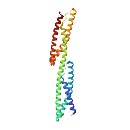
    1S35

  • PubMed Abstract: 

    Erythroid spectrin, a major component of the cytoskeletal network of the red cell which contributes to both the stability and the elasticity of the red cell membrane, is composed of two subunits, alpha and beta, each formed by 16-20 tandem repeats. The properties of the repeats and their relative arrangement are thought to be key determinants of spectrin flexibility. Here we report a 2.4 A resolution crystal structure of human erythroid beta-spectrin repeats 8 and 9. This two-repeat fragment is unusual as it exhibits low stability of folding and one of its repeats lacks two tryptophans highly conserved among spectrin repeats. Two key factors responsible for the lower stability and, possibly, its flexibility, are revealed by the structure. A third novel feature of the structure is the relative orientation of the two repeats, which increases the range of possible conformations and provides new insights into atomic models of spectrin flexibility.


  • Organizational Affiliation

    Department of Biochemistry, Molecular Biology, and Cell Biology, Northwestern University, 2205 Tech Drive, Evanston, IL 60208 USA.


Macromolecules
Find similar proteins by:  (by identity cutoff)  |  3D Structure
Entity ID: 1
MoleculeChains Sequence LengthOrganismDetailsImage
Spectrin beta chain, erythrocyte214Homo sapiensMutation(s): 1 
Gene Names: SPTBSPTB1
UniProt & NIH Common Fund Data Resources
Find proteins for P11277 (Homo sapiens)
Explore P11277 
Go to UniProtKB:  P11277
PHAROS:  P11277
GTEx:  ENSG00000070182 
Entity Groups  
Sequence Clusters30% Identity50% Identity70% Identity90% Identity95% Identity100% Identity
UniProt GroupP11277
Sequence Annotations
Expand
  • Reference Sequence
Experimental Data & Validation

Experimental Data

  • Method: X-RAY DIFFRACTION
  • Resolution: 2.40 Å
  • R-Value Free: 0.259 
  • R-Value Work: 0.222 
  • Space Group: P 41 21 2
Unit Cell:
Length ( Å )Angle ( ˚ )
a = 122.151α = 90
b = 122.151β = 90
c = 49.548γ = 90
Software Package:
Software NamePurpose
XDSdata scaling
SCALAdata scaling
SHARPphasing
SOLVEphasing
CNSrefinement
XDSdata reduction
CCP4data scaling

Structure Validation

View Full Validation Report


Entry History 

Deposition Data

Revision History  (Full details and data files)

  • Version 1.0: 2004-04-20
    Type: Initial release
  • Version 1.1: 2008-04-29
    Changes: Version format compliance
  • Version 1.2: 2011-07-13
    Changes: Version format compliance
  • Version 1.3: 2021-10-27
    Changes: Database references, Derived calculations
  • Version 1.4: 2024-02-14
    Changes: Data collection