1RZY

Crystal structure of rabbit Hint complexed with N-ethylsulfamoyladenosine


Experimental Data Snapshot

  • Method: X-RAY DIFFRACTION
  • Resolution: 1.80 Å
  • R-Value Free: 0.242 
  • R-Value Work: 0.216 
  • R-Value Observed: 0.217 

wwPDB Validation   3D Report Full Report


Ligand Structure Quality Assessment 


This is version 1.3 of the entry. See complete history


Literature

Biochemical, crystallographic, and mutagenic characterization of hint, the AMP-lysine hydrolase, with novel substrates and inhibitors

Krakowiak, A.K.Pace, H.C.Blackburn, G.M.Adams, M.Mekhalfia, A.Kaczmarek, R.Baraniak, J.Stec, W.J.Brenner, C.

(2004) J Biol Chem 279: 18711-18716

  • DOI: https://doi.org/10.1074/jbc.M314271200
  • Primary Citation of Related Structures:  
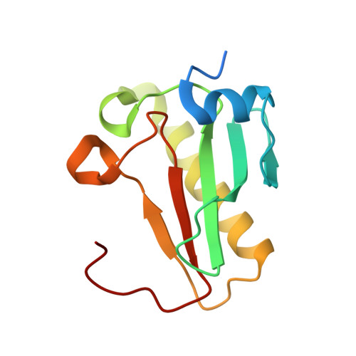
    1RZY

  • PubMed Abstract: 

    Hint, histidine triad nucleotide-binding protein, is a universally conserved enzyme that hydrolyzes AMP linked to lysine and, in yeast, functions as a positive regulator of the RNA polymerase II C-terminal domain kinase, Kin28. To explore the biochemical and structural bases for the adenosine phosphoramidate hydrolase activity of rabbit Hint, we synthesized novel substrates linking a p-nitroaniline group to adenylate (AMP-pNA) and inhibitors that consist of an adenosine group and 5'-sulfamoyl (AdoOSO(2)NH(2)) or N-ethylsulfamoyl (AdoOSO(2)NHCH(2)CH(3)) group. AMP-pNA is a suitable substrate for Hint that allowed characterization of the inhibitors; titration of each inhibitor into AMP-pNA assays revealed their K(i) values. The N-ethylsulfamoyl derivative has a 13-fold binding advantage over the sulfamoyl adenosine. The 1.8-A cocrystal structure of rabbit Hint with N-ethylsulfamoyl adenosine revealed a binding site for the ethyl group against Trp-123, a residue that reaches across the Hint dimer interface to interact with the alkyl portion of the inhibitor and, presumably, the alkyl portion of a lysyl substrate. Ser-107 is positioned to donate a hydrogen bond to the leaving group nitrogen. Consistent with a role in acid-base catalysis, the Hint S107A mutant protein displayed depressed catalytic activity.


  • Organizational Affiliation

    Structural Biology and Bioinformatics Program, Kimmel Cancer Center, Philadelphia, Pennsylvania 19107, USA.


Macromolecules
Find similar proteins by:  (by identity cutoff)  |  3D Structure
Entity ID: 1
MoleculeChains Sequence LengthOrganismDetailsImage
Histidine triad nucleotide-binding protein 1126Oryctolagus cuniculusMutation(s): 0 
Gene Names: HINT1HINT
UniProt
Find proteins for P80912 (Oryctolagus cuniculus)
Explore P80912 
Go to UniProtKB:  P80912
Entity Groups  
Sequence Clusters30% Identity50% Identity70% Identity90% Identity95% Identity100% Identity
UniProt GroupP80912
Sequence Annotations
Expand
  • Reference Sequence
Small Molecules
Ligands 1 Unique
IDChains Name / Formula / InChI Key2D Diagram3D Interactions
5AS
Query on 5AS

Download Ideal Coordinates CCD File 
B [auth A]5'-O-(N-ETHYL-SULFAMOYL)ADENOSINE
C12 H18 N6 O6 S
ONCSLXAPOGUODU-WOUKDFQISA-N
Binding Affinity Annotations 
IDSourceBinding Affinity
5AS Binding MOAD:  1RZY Ki: 1250 (nM) from 1 assay(s)
Experimental Data & Validation

Experimental Data

  • Method: X-RAY DIFFRACTION
  • Resolution: 1.80 Å
  • R-Value Free: 0.242 
  • R-Value Work: 0.216 
  • R-Value Observed: 0.217 
  • Space Group: P 43 21 2
Unit Cell:
Length ( Å )Angle ( ˚ )
a = 39.888α = 90
b = 39.888β = 90
c = 142.286γ = 90
Software Package:
Software NamePurpose
crystaldata collection
d*TREKdata reduction
CNSrefinement
CrystalCleardata reduction
d*TREKdata scaling
CNSphasing

Structure Validation

View Full Validation Report


Ligand Structure Quality Assessment 


Entry History 

Deposition Data

Revision History  (Full details and data files)

  • Version 1.0: 2004-03-09
    Type: Initial release
  • Version 1.1: 2008-04-29
    Changes: Version format compliance
  • Version 1.2: 2011-07-13
    Changes: Derived calculations, Version format compliance
  • Version 1.3: 2024-02-14
    Changes: Data collection, Database references, Derived calculations