Backbone solution structures of proteins using residual dipolar couplings: application to a novel structural genomics target.
Valafar, H., Mayer, K.L., Bougault, C.M., LeBlond, P.D., Jenney, F.E., Brereton, P.S., Adams, M.W., Prestegard, J.H.(2004) J Struct Funct Genomics 5: 241-254
- PubMed: 15704012
- DOI: https://doi.org/10.1007/s10969-005-4899-5
- Primary Citation of Related Structures:
1RWS, 1SF0 - PubMed Abstract:
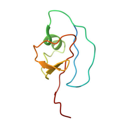
Structural genomics (or proteomics) activities are critically dependent on the availability of high-throughput structure determination methodology. Development of such methodology has been a particular challenge for NMR based structure determination because of the demands for isotopic labeling of proteins and the requirements for very long data acquisition times. We present here a methodology that gains efficiency from a focus on determination of backbone structures of proteins as opposed to full structures with all sidechains in place. This focus is appropriate given the presumption that many protein structures in the future will be built using computational methods that start from representative fold family structures and replace as many as 70% of the sidechains in the course of structure determination. The methodology we present is based primarily on residual dipolar couplings (RDCs), readily accessible NMR observables that constrain the orientation of backbone fragments irrespective of separation in space. A new software tool is described for the assembly of backbone fragments under RDC constraints and an application to a structural genomics target is presented. The target is an 8.7 kDa protein from Pyrococcus furiosus, PF1061, that was previously not well annotated, and had a nearest structurally characterized neighbor with only 33% sequence identity. The structure produced shows structural similarity to this sequence homologue, but also shows similarity to other proteins, which suggests a functional role in sulfur transfer. Given the backbone structure and a possible functional link this should be an ideal target for development of modeling methods.
Organizational Affiliation:
Southeast Collaboratory for Structural Genomics, University of Georgia, Athens, GA 30602, USA.