1RV1

CRYSTAL STRUCTURE OF HUMAN MDM2 WITH AN IMIDAZOLINE INHIBITOR


Experimental Data Snapshot

  • Method: X-RAY DIFFRACTION
  • Resolution: 2.30 Å
  • R-Value Free: 0.322 
  • R-Value Work: 0.256 
  • R-Value Observed: 0.266 

wwPDB Validation   3D Report Full Report


This is version 1.4 of the entry. See complete history


Literature

In vivo activation of the p53 pathway by small-molecule antagonists of MDM2.

Vassilev, L.T.Vu, B.T.Graves, B.Carvajal, D.Podlaski, F.Filipovic, Z.Kong, N.Kammlott, U.Lukacs, C.Klein, C.Fotouhi, N.Liu, E.A.

(2004) Science 303: 844-848

  • DOI: https://doi.org/10.1126/science.1092472
  • Primary Citation of Related Structures:  
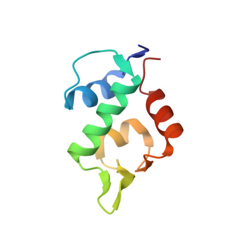
    1RV1

  • PubMed Abstract: 

    MDM2 binds the p53 tumor suppressor protein with high affinity and negatively modulates its transcriptional activity and stability. Overexpression of MDM2, found in many human tumors, effectively impairs p53 function. Inhibition of MDM2-p53 interaction can stabilize p53 and may offer a novel strategy for cancer therapy. Here, we identify potent and selective small-molecule antagonists of MDM2 and confirm their mode of action through the crystal structures of complexes. These compounds bind MDM2 in the p53-binding pocket and activate the p53 pathway in cancer cells, leading to cell cycle arrest, apoptosis, and growth inhibition of human tumor xenografts in nude mice.


  • Organizational Affiliation

    Department of Discovery Oncology, Roche Research Center, Hoffmann-La Roche, Inc., Nutley, NJ 07110, USA. lyubomir.vassilev@roche.com


Macromolecules
Find similar proteins by:  (by identity cutoff)  |  3D Structure
Entity ID: 1
MoleculeChains Sequence LengthOrganismDetailsImage
Ubiquitin-protein ligase E3 Mdm2
A, B, C
85Homo sapiensMutation(s): 1 
Gene Names: MDM2
EC: 6.3.2
UniProt & NIH Common Fund Data Resources
Find proteins for Q00987 (Homo sapiens)
Explore Q00987 
Go to UniProtKB:  Q00987
PHAROS:  Q00987
GTEx:  ENSG00000135679 
Entity Groups  
Sequence Clusters30% Identity50% Identity70% Identity90% Identity95% Identity100% Identity
UniProt GroupQ00987
Sequence Annotations
Expand
  • Reference Sequence
Small Molecules
Ligands 1 Unique
IDChains Name / Formula / InChI Key2D Diagram3D Interactions
IMZ
Query on IMZ

Download Ideal Coordinates CCD File 
D [auth A],
E [auth A],
F [auth B],
G [auth B],
H [auth C]
CIS-[4,5-BIS-(4-BROMOPHENYL)-2-(2-ETHOXY-4-METHOXYPHENYL)-4,5-DIHYDROIMIDAZOL-1-YL]-[4-(2-HYDROXYETHYL)PIPERAZIN-1-YL]METHANONE
C31 H34 Br2 N4 O4
PVRYEWOXWGDQHA-URLMMPGGSA-N
Binding Affinity Annotations 
IDSourceBinding Affinity
IMZ BindingDB:  1RV1 IC50: min: 100, max: 140 (nM) from 2 assay(s)
Binding MOAD:  1RV1 IC50: 140 (nM) from 1 assay(s)
PDBBind:  1RV1 IC50: 140 (nM) from 1 assay(s)
Experimental Data & Validation

Experimental Data

  • Method: X-RAY DIFFRACTION
  • Resolution: 2.30 Å
  • R-Value Free: 0.322 
  • R-Value Work: 0.256 
  • R-Value Observed: 0.266 
  • Space Group: C 1 2 1
Unit Cell:
Length ( Å )Angle ( ˚ )
a = 122.264α = 90
b = 39.727β = 114.29
c = 79.362γ = 90
Software Package:
Software NamePurpose
HKL-2000data collection
SCALEPACKdata scaling
MOLREPphasing
CNXrefinement
HKL-2000data reduction

Structure Validation

View Full Validation Report


Entry History 

Deposition Data

Revision History  (Full details and data files)

  • Version 1.0: 2004-01-20
    Type: Initial release
  • Version 1.1: 2008-04-29
    Changes: Version format compliance
  • Version 1.2: 2011-07-13
    Changes: Version format compliance
  • Version 1.3: 2021-10-27
    Changes: Database references, Derived calculations
  • Version 1.4: 2023-08-23
    Changes: Data collection, Refinement description