1RUS

CRYSTAL STRUCTURE OF THE BINARY COMPLEX OF RIBULOSE-1,5-BISPHOSPHATE CARBOXYLASE AND ITS PRODUCT, 3-PHOSPHO-D-GLYCERATE


Experimental Data Snapshot

  • Method: X-RAY DIFFRACTION
  • Resolution: 2.90 Å
  • R-Value Observed: 0.203 

wwPDB Validation   3D Report Full Report


This is version 1.4 of the entry. See complete history


Literature

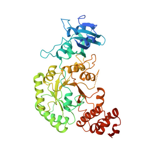
Crystal structure of the binary complex of ribulose-1,5-bisphosphate carboxylase and its product, 3-phospho-D-glycerate.

Lundqvist, T.Schneider, G.

(1989) J Biol Chem 264: 3643-3646

  • DOI: https://doi.org/10.2210/pdb1rus/pdb
  • Primary Citation of Related Structures:  
    1RUS

  • PubMed Abstract: 

    The crystal structure of the binary complex of non-activated ribulose-1,5-bisphosphate carboxylase/oxygenase from Rhodospirillum rubrum and its product 3-phospho-D-glycerate has been determined to 2.9-A resolution. This structure determination confirms the proposed location of the active site (Schneider, G., Lindqvist, Y., Brändén, C.-I., and Lorimer, G. (1986) EMBO J. 5, 3409-3415) at the carboxyl end of the beta-strands of the alpha/beta-barrel in the carboxyl-terminal domain. One molecule of 3-phosphoglycerate is bound per active site. All oxygen atoms of 3-phosphoglycerate form hydrogen bonds to groups of the enzyme. The phosphate group interacts with the sidechains of residues Arg-288, His-321, and Ser-368, which are conserved between enzymes from different species as well as with the main chain nitrogens from residues Thr-322 and Gly-323. These amino acid residues constitute one of the two phosphate binding sites of the active site. The carboxyl group interacts with the side chains of His-287, Lys-191, and Asn-111. Implications of the activation process for the binding of 3-phosphoglycerate are discussed.


  • Organizational Affiliation

    Swedish University of Agricultural Sciences, Department of Molecular Biology, Uppsala Biomedical Center, Sweden.


Macromolecules
Find similar proteins by:  (by identity cutoff)  |  3D Structure
Entity ID: 1
MoleculeChains Sequence LengthOrganismDetailsImage
RUBISCO (RIBULOSE-1,5-BISPHOSPHATE CARBOXYLASE(SLASH)OXYGENASE)
A, B
490Rhodospirillum rubrumMutation(s): 0 
EC: 4.1.1.39
UniProt
Find proteins for P04718 (Rhodospirillum rubrum)
Explore P04718 
Go to UniProtKB:  P04718
Entity Groups  
Sequence Clusters30% Identity50% Identity70% Identity90% Identity95% Identity100% Identity
UniProt GroupP04718
Sequence Annotations
Expand
  • Reference Sequence
Small Molecules
Ligands 1 Unique
IDChains Name / Formula / InChI Key2D Diagram3D Interactions
3PG
Query on 3PG

Download Ideal Coordinates CCD File 
C [auth A],
D [auth B]
3-PHOSPHOGLYCERIC ACID
C3 H7 O7 P
OSJPPGNTCRNQQC-UWTATZPHSA-N
Experimental Data & Validation

Experimental Data

  • Method: X-RAY DIFFRACTION
  • Resolution: 2.90 Å
  • R-Value Observed: 0.203 
  • Space Group: P 1 21 1
Unit Cell:
Length ( Å )Angle ( ˚ )
a = 65.5α = 90
b = 70.6β = 92.1
c = 104.1γ = 90
Software Package:
Software NamePurpose
PROLSQrefinement

Structure Validation

View Full Validation Report


Entry History 

Deposition Data

Revision History  (Full details and data files)

  • Version 1.0: 1991-10-15
    Type: Initial release
  • Version 1.1: 2008-03-24
    Changes: Version format compliance
  • Version 1.2: 2011-07-13
    Changes: Version format compliance
  • Version 1.3: 2017-11-29
    Changes: Advisory, Derived calculations, Other
  • Version 1.4: 2024-02-14
    Changes: Advisory, Data collection, Database references, Derived calculations