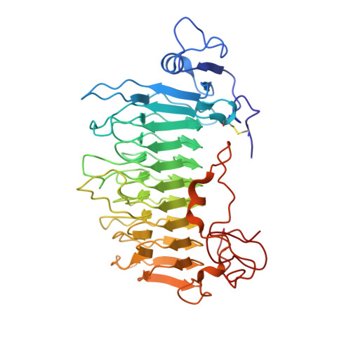
The crystal structure of pectate lyase Pel9A from Erwinia chrysanthemi
Jenkins, J., Shevchik, V.E., Hugouvieux-Cotte-Pattat, N., Pickersgill, R.W.(2004) J Biol Chem 279: 9139-9145
- PubMed: 14670977
- DOI: https://doi.org/10.1074/jbc.M311390200
- Primary Citation of Related Structures:
1RU4 - PubMed Abstract:
The "family 9 polysaccharide lyase" pectate lyase L (Pel9A) from Erwinia chrysanthemi comprises a 10-coil parallel beta-helix domain with distinct structural features including an asparagine ladder and aromatic stack at novel positions within the superhelical structure. Pel9A has a single high affinity calcium-binding site strikingly similar to the "primary" calcium-binding site described previously for the family Pel1A pectate lyases, and there is strong evidence for a common second calcium ion that binds between enzyme and substrate in the "Michaelis" complex. Although the primary calcium ion binds substrate in subsite -1, it is the second calcium ion, whose binding site is formed by the coming together of enzyme and substrate, that facilitates abstraction of the C5 proton from the sacharride in subsite +1. The role of the second calcium is to withdraw electrons from the C6 carboxylate of the substrate, thereby acidifying the C5 proton facilitating its abstraction and resulting in an E1cb-like anti-beta-elimination mechanism. The active site geometries and mechanism of Pel1A and Pel9A are closely similar, but the catalytic base is a lysine in the Pel9A enzymes as opposed to an arginine in the Pel1A enzymes.
Organizational Affiliation:
Institute of Food Research, Norwich Research Park, Colney Lane, Norwich, NR4 7UA, United Kingdom.