1RTM

TRIMERIC STRUCTURE OF A C-TYPE MANNOSE-BINDING PROTEIN


Experimental Data Snapshot

  • Method: X-RAY DIFFRACTION
  • Resolution: 1.80 Å
  • R-Value Free: 0.270 
  • R-Value Work: 0.220 
  • R-Value Observed: 0.220 

wwPDB Validation   3D Report Full Report


This is version 1.2 of the entry. See complete history


Literature

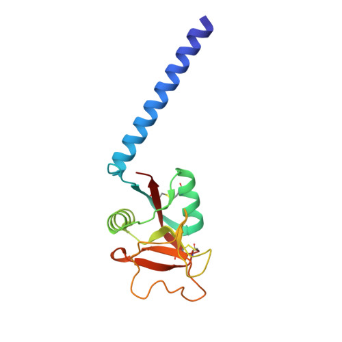
Trimeric structure of a C-type mannose-binding protein.

Weis, W.I.Drickamer, K.

(1994) Structure 2: 1227-1240

  • DOI: https://doi.org/10.1016/S0969-2126(94)00124-3
  • Primary Citation of Related Structures:  
    1RTM

  • PubMed Abstract: 

    Mannose-binding proteins (MBPs) are C-type (Ca(2+)-dependent) animal lectins found in serum. They recognize cell-surface oligosaccharide structures characteristic of pathogenic bacteria and fungi, and trigger the neutralization of these organisms. Like most lectins, MBPs display weak intrinsic affinity for monovalent sugar ligands, but bind avidly to multivalent ligands.


  • Organizational Affiliation

    Department of Structural Biology, Stanford University School of Medicine, CA 94305.


Macromolecules
Find similar proteins by:  (by identity cutoff)  |  3D Structure
Entity ID: 1
MoleculeChains Sequence LengthOrganismDetailsImage
MANNOSE-BINDING PROTEIN-AA [auth 1],
B [auth 2],
C [auth 3]
149Rattus norvegicusMutation(s): 0 
UniProt
Find proteins for P19999 (Rattus norvegicus)
Explore P19999 
Go to UniProtKB:  P19999
Entity Groups  
Sequence Clusters30% Identity50% Identity70% Identity90% Identity95% Identity100% Identity
UniProt GroupP19999
Sequence Annotations
Expand
  • Reference Sequence
Small Molecules
Ligands 3 Unique
IDChains Name / Formula / InChI Key2D Diagram3D Interactions
GOL
Query on GOL

Download Ideal Coordinates CCD File 
BA [auth 3]
CA [auth 3]
DA [auth 3]
H [auth 1]
I [auth 1]
BA [auth 3],
CA [auth 3],
DA [auth 3],
H [auth 1],
I [auth 1],
J [auth 1],
K [auth 1],
L [auth 1],
M [auth 1],
N [auth 1],
O [auth 1],
P [auth 1],
U [auth 2],
V [auth 2],
W [auth 2]
GLYCEROL
C3 H8 O3
PEDCQBHIVMGVHV-UHFFFAOYSA-N
CA
Query on CA

Download Ideal Coordinates CCD File 
D [auth 1]
E [auth 1]
F [auth 1]
Q [auth 2]
R [auth 2]
D [auth 1],
E [auth 1],
F [auth 1],
Q [auth 2],
R [auth 2],
S [auth 2],
X [auth 3],
Y [auth 3],
Z [auth 3]
CALCIUM ION
Ca
BHPQYMZQTOCNFJ-UHFFFAOYSA-N
CL
Query on CL

Download Ideal Coordinates CCD File 
AA [auth 3],
G [auth 1],
T [auth 2]
CHLORIDE ION
Cl
VEXZGXHMUGYJMC-UHFFFAOYSA-M
Experimental Data & Validation

Experimental Data

  • Method: X-RAY DIFFRACTION
  • Resolution: 1.80 Å
  • R-Value Free: 0.270 
  • R-Value Work: 0.220 
  • R-Value Observed: 0.220 
  • Space Group: C 1 2 1
Unit Cell:
Length ( Å )Angle ( ˚ )
a = 80α = 90
b = 85.1β = 106.3
c = 98.5γ = 90
Software Package:
Software NamePurpose
X-PLORmodel building
X-PLORrefinement
X-PLORphasing

Structure Validation

View Full Validation Report


Entry History 

Deposition Data

Revision History  (Full details and data files)

  • Version 1.0: 1995-02-07
    Type: Initial release
  • Version 1.1: 2008-03-03
    Changes: Version format compliance
  • Version 1.2: 2011-07-13
    Changes: Non-polymer description, Version format compliance