1RSY

STRUCTURE OF THE FIRST C2-DOMAIN OF SYNAPTOTAGMIN I: A NOVEL CA2+(SLASH)PHOSPHOLIPID BINDING FOLD


Experimental Data Snapshot

  • Method: X-RAY DIFFRACTION
  • Resolution: 1.90 Å
  • R-Value Free: 0.249 
  • R-Value Work: 0.186 
  • R-Value Observed: 0.186 

wwPDB Validation   3D Report Full Report


This is version 1.3 of the entry. See complete history

Re-refinement Note

A newer entry is available that reflects an alternative modeling of the original data: 4WEE


Literature

Structure of the first C2 domain of synaptotagmin I: a novel Ca2+/phospholipid-binding fold.

Sutton, R.B.Davletov, B.A.Berghuis, A.M.Sudhof, T.C.Sprang, S.R.

(1995) Cell 80: 929-938

  • DOI: https://doi.org/10.1016/0092-8674(95)90296-1
  • Primary Citation of Related Structures:  
    1RSY

  • PubMed Abstract: 

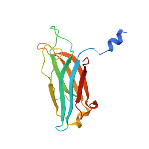
    C2 domains are regulatory sequence motifs that occur widely in nature. Synaptotagmin I, a synaptic vesicle protein involved in the Ca2+ regulation of exocytosis, contains two C2 domains, the first of which acts as a Ca2+ sensor. We now describe the three-dimensional structure of this C2 domain at 1.9 A resolution in both the Ca(2+)-bound and Ca(2+)-free forms. The C2 polypeptide forms an eight-stranded beta sandwich constructed around a conserved four-stranded motif designated as a C2 key. Ca2+ binds in a cup-shaped depression between two polypeptide loops located at the N- and C-termini of the C2-key motif.


  • Organizational Affiliation

    Department of Biochemistry, University of Texas Southwestern Medical Center, Dallas 75235.


Macromolecules
Find similar proteins by:  (by identity cutoff)  |  3D Structure
Entity ID: 1
MoleculeChains Sequence LengthOrganismDetailsImage
SYNAPTOTAGMIN I152Rattus norvegicusMutation(s): 0 
Membrane Entity: Yes 
UniProt
Find proteins for P21707 (Rattus norvegicus)
Explore P21707 
Go to UniProtKB:  P21707
Entity Groups  
Sequence Clusters30% Identity50% Identity70% Identity90% Identity95% Identity100% Identity
UniProt GroupP21707
Sequence Annotations
Expand
  • Reference Sequence
Small Molecules
Ligands 1 Unique
IDChains Name / Formula / InChI Key2D Diagram3D Interactions
SO4
Query on SO4

Download Ideal Coordinates CCD File 
B [auth A]SULFATE ION
O4 S
QAOWNCQODCNURD-UHFFFAOYSA-L
Experimental Data & Validation

Experimental Data

  • Method: X-RAY DIFFRACTION
  • Resolution: 1.90 Å
  • R-Value Free: 0.249 
  • R-Value Work: 0.186 
  • R-Value Observed: 0.186 
  • Space Group: P 1 21 1
Unit Cell:
Length ( Å )Angle ( ˚ )
a = 42.3α = 90
b = 38.9β = 96.9
c = 44.6γ = 90
Software Package:
Software NamePurpose
X-PLORmodel building
X-PLORrefinement
DENZOdata reduction
X-PLORphasing

Structure Validation

View Full Validation Report


Entry History 

Deposition Data

Revision History  (Full details and data files)

  • Version 1.0: 1995-05-08
    Type: Initial release
  • Version 1.1: 2008-03-03
    Changes: Version format compliance
  • Version 1.2: 2011-07-13
    Changes: Version format compliance
  • Version 1.3: 2024-02-14
    Changes: Data collection, Database references, Derived calculations, Other