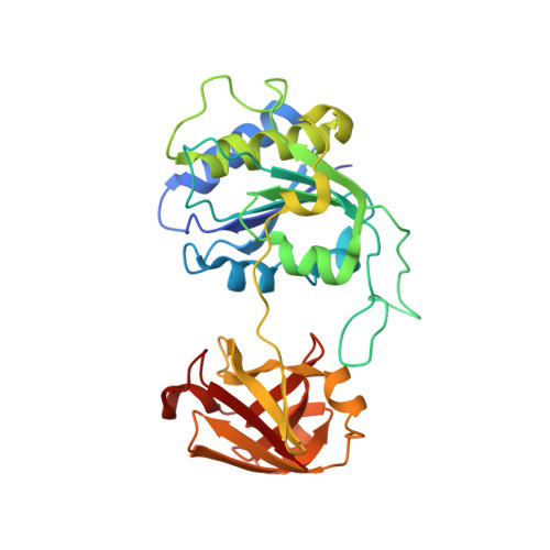
Crystal structure and mechanism of a bacterial fluorinating enzyme
Dong, C., Huang, F., Deng, H., Schaffrath, C., Spencer, J.B., O'Hagan, D., Naismith, J.H.(2004) Nature 427: 561-565
- PubMed: 14765200
- DOI: https://doi.org/10.1038/nature02280
- Primary Citation of Related Structures:
1RQP, 1RQR - PubMed Abstract:
Fluorine is the thirteenth most abundant element in the earth's crust, but fluoride concentrations in surface water are low and fluorinated metabolites are extremely rare. The fluoride ion is a potent nucleophile in its desolvated state, but is tightly hydrated in water and effectively inert. Low availability and a lack of chemical reactivity have largely excluded fluoride from biochemistry: in particular, fluorine's high redox potential precludes the haloperoxidase-type mechanism used in the metabolic incorporation of chloride and bromide ions. But fluorinated chemicals are growing in industrial importance, with applications in pharmaceuticals, agrochemicals and materials products. Reactive fluorination reagents requiring specialist process technologies are needed in industry and, although biological catalysts for these processes are highly sought after, only one enzyme that can convert fluoride to organic fluorine has been described. Streptomyces cattleya can form carbon-fluorine bonds and must therefore have evolved an enzyme able to overcome the chemical challenges of using aqueous fluoride. Here we report the sequence and three-dimensional structure of the first native fluorination enzyme, 5'-fluoro-5'-deoxyadenosine synthase, from this organism. Both substrate and products have been observed bound to the enzyme, enabling us to propose a nucleophilic substitution mechanism for this biological fluorination reaction.
Organizational Affiliation:
Centre for Biomolecular Sciences, The University of St Andrews, Fife KY16 9ST, UK.