1RP4

Structure of Ero1p, Source of Disulfide Bonds for Oxidative Protein Folding in the Cell


Experimental Data Snapshot

  • Method: X-RAY DIFFRACTION
  • Resolution: 2.20 Å
  • R-Value Free: 0.244 
  • R-Value Work: 0.217 

wwPDB Validation   3D Report Full Report


Ligand Structure Quality Assessment 


This is version 1.3 of the entry. See complete history


Literature

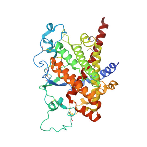
Structure of ero1p, source of disulfide bonds for oxidative protein folding in the cell.

Gross, E.Kastner, D.B.Kaiser, C.A.Fass, D.

(2004) Cell 117: 601-610

  • DOI: https://doi.org/10.1016/s0092-8674(04)00418-0
  • Primary Citation of Related Structures:  
    1RP4, 1RQ1

  • PubMed Abstract: 

    The flavoenzyme Ero1p produces disulfide bonds for oxidative protein folding in the endoplasmic reticulum. Disulfides generated de novo within Ero1p are transferred to protein disulfide isomerase and then to substrate proteins by dithiol-disulfide exchange reactions. Despite this key role of Ero1p, little is known about the mechanism by which this enzyme catalyzes thiol oxidation. Here, we present the X-ray crystallographic structure of Ero1p, which reveals the molecular details of the catalytic center, the role of a CXXCXXC motif, and the spatial relationship between functionally significant cysteines and the bound cofactor. Remarkably, the Ero1p active site closely resembles that of the versatile thiol oxidase module of Erv2p, a protein with no sequence homology to Ero1p. Furthermore, both Ero1p and Erv2p display essential dicysteine motifs on mobile polypeptide segments, suggesting that shuttling electrons to a rigid active site using a flexible strand is a fundamental feature of disulfide-generating flavoenzymes.


  • Organizational Affiliation

    Department of Structural Biology, Weizmann Institute of Science, Rehovot 76100, Israel.


Macromolecules
Find similar proteins by:  (by identity cutoff)  |  3D Structure
Entity ID: 1
MoleculeChains Sequence LengthOrganismDetailsImage
Hypothetical 65.0 kDa protein in COX14-COS3 intergenic region precursor389Saccharomyces cerevisiaeMutation(s): 7 
Gene Names: YML130CYM4987.05C
Membrane Entity: Yes 
UniProt
Find proteins for Q03103 (Saccharomyces cerevisiae (strain ATCC 204508 / S288c))
Explore Q03103 
Go to UniProtKB:  Q03103
Entity Groups  
Sequence Clusters30% Identity50% Identity70% Identity90% Identity95% Identity100% Identity
UniProt GroupQ03103
Sequence Annotations
Expand
  • Reference Sequence
Small Molecules
Modified Residues  1 Unique
IDChains TypeFormula2D DiagramParent
MSE
Query on MSE
A
L-PEPTIDE LINKINGC5 H11 N O2 SeMET
Experimental Data & Validation

Experimental Data

  • Method: X-RAY DIFFRACTION
  • Resolution: 2.20 Å
  • R-Value Free: 0.244 
  • R-Value Work: 0.217 
  • Space Group: C 2 2 21
Unit Cell:
Length ( Å )Angle ( ˚ )
a = 73.47α = 90
b = 134.33β = 90
c = 102.68γ = 90
Software Package:
Software NamePurpose
DENZOdata reduction
SCALEPACKdata scaling
SHARPphasing
CNSrefinement

Structure Validation

View Full Validation Report


Ligand Structure Quality Assessment 


Entry History 

Deposition Data

Revision History  (Full details and data files)

  • Version 1.0: 2004-06-08
    Type: Initial release
  • Version 1.1: 2008-04-29
    Changes: Version format compliance
  • Version 1.2: 2011-07-13
    Changes: Version format compliance
  • Version 1.3: 2018-01-31
    Changes: Experimental preparation