1RL6

RIBOSOMAL PROTEIN L6


Experimental Data Snapshot

  • Method: X-RAY DIFFRACTION
  • Resolution: 2.00 Å
  • R-Value Free: 0.302 
  • R-Value Work: 0.257 

wwPDB Validation   3D Report Full Report


This is version 1.4 of the entry. See complete history


Literature

Ribosomal protein L6: structural evidence of gene duplication from a primitive RNA binding protein.

Golden, B.L.Ramakrishnan, V.White, S.W.

(1993) EMBO J 12: 4901-4908

  • DOI: https://doi.org/10.1002/j.1460-2075.1993.tb06184.x
  • Primary Citation of Related Structures:  
    1RL6

  • PubMed Abstract: 

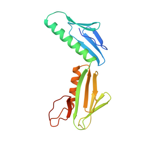
    In all cells, protein synthesis is coordinated by the ribosome, a large ribonucleoprotein particle that is composed of > 50 distinct protein molecules and several large RNA molecules. Here we present the crystal structure of ribosomal protein L6 from the thermophilic bacterium Bacillus stearothermophilus solved at 2.6 A resolution. L6 contains two domains with almost identical folds, implying that it was created by an ancient gene duplication event. The surface of the molecule displays several likely sites of interaction with other components of the ribosome. The RNA binding sites appear to be localized in the C-terminal domain whereas the N-terminal domain contains the potential sites for protein-protein interactions. The domain structure is homologous with several other ribosomal proteins and to a large family of eukaryotic RNA binding proteins.


  • Organizational Affiliation

    Department of Microbiology, Duke University Medical Center, Durham, NC 27710.


Macromolecules
Find similar proteins by:  (by identity cutoff)  |  3D Structure
Entity ID: 1
MoleculeChains Sequence LengthOrganismDetailsImage
PROTEIN (RIBOSOMAL PROTEIN L6)177Geobacillus stearothermophilusMutation(s): 0 
UniProt
Find proteins for P02391 (Geobacillus stearothermophilus)
Explore P02391 
Go to UniProtKB:  P02391
Entity Groups  
Sequence Clusters30% Identity50% Identity70% Identity90% Identity95% Identity100% Identity
UniProt GroupP02391
Sequence Annotations
Expand
  • Reference Sequence
Experimental Data & Validation

Experimental Data

  • Method: X-RAY DIFFRACTION
  • Resolution: 2.00 Å
  • R-Value Free: 0.302 
  • R-Value Work: 0.257 
  • Space Group: P 61 2 2
Unit Cell:
Length ( Å )Angle ( ˚ )
a = 71.8α = 90
b = 71.8β = 90
c = 124.5γ = 120
Software Package:
Software NamePurpose
PHASESphasing
X-PLORrefinement
DENZOdata reduction
SCALEPACKdata scaling

Structure Validation

View Full Validation Report


Entry History 

Deposition Data

Revision History  (Full details and data files)

  • Version 1.0: 1999-02-02
    Type: Initial release
  • Version 1.1: 2008-04-26
    Changes: Version format compliance
  • Version 1.2: 2011-07-13
    Changes: Version format compliance
  • Version 1.3: 2019-10-30
    Changes: Data collection, Database references, Structure summary
  • Version 1.4: 2023-12-27
    Changes: Data collection, Database references