1RKC

Human vinculin head (1-258) in complex with talin's vinculin binding site 3 (residues 1944-1969)


Experimental Data Snapshot

  • Method: X-RAY DIFFRACTION
  • Resolution: 2.70 Å
  • R-Value Free: 0.335 
  • R-Value Work: 0.244 

wwPDB Validation   3D Report Full Report


This is version 1.3 of the entry. See complete history


Literature

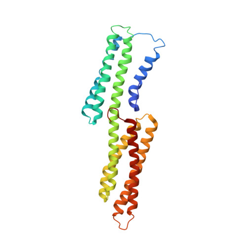
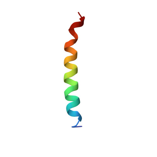
Vinculin activation by talin through helical bundle conversion

Izard, T.Evans, G.Borgon, R.A.Rush, C.L.Bricogne, G.Bois, P.R.

(2004) Nature 427: 171-175

  • DOI: https://doi.org/10.1038/nature02281
  • Primary Citation of Related Structures:  
    1RKC, 1RKE

  • PubMed Abstract: 

    Vinculin is a conserved component and an essential regulator of both cell-cell (cadherin-mediated) and cell-matrix (integrin-talin-mediated focal adhesions) junctions, and it anchors these adhesion complexes to the actin cytoskeleton by binding to talin in integrin complexes or to alpha-actinin in cadherin junctions. In its resting state, vinculin is held in a closed conformation through interactions between its head (Vh) and tail (Vt) domains. The binding of vinculin to focal adhesions requires its association with talin. Here we report the crystal structures of human vinculin in its inactive and talin-activated states. Talin binding induces marked conformational changes in Vh, creating a novel helical bundle structure, and this alteration actively displaces Vt from Vh. These results, as well as the ability of alpha-actinin to also bind to Vh and displace Vt from pre-existing Vh-Vt complexes, support a model whereby Vh functions as a domain that undergoes marked structural changes that allow vinculin to direct cytoskeletal assembly in focal adhesions and adherens junctions. Notably, talin's effects on Vh structure establish helical bundle conversion as a signalling mechanism by which proteins direct cellular responses.


  • Organizational Affiliation

    Department of Hematology-Oncology, St Jude Children's Research Hospital, Memphis, Tennessee 38105, USA. tina.izard@stjude.org


Macromolecules
Find similar proteins by:  (by identity cutoff)  |  3D Structure
Entity ID: 1
MoleculeChains Sequence LengthOrganismDetailsImage
Vinculin262Homo sapiensMutation(s): 0 
Gene Names: VCL
UniProt & NIH Common Fund Data Resources
Find proteins for P18206 (Homo sapiens)
Explore P18206 
Go to UniProtKB:  P18206
PHAROS:  P18206
GTEx:  ENSG00000035403 
Entity Groups  
Sequence Clusters30% Identity50% Identity70% Identity90% Identity95% Identity100% Identity
UniProt GroupP18206
Sequence Annotations
Expand
  • Reference Sequence
Find similar proteins by:  (by identity cutoff)  |  3D Structure
Entity ID: 2
MoleculeChains Sequence LengthOrganismDetailsImage
Talin26Gallus gallusMutation(s): 0 
UniProt
Find proteins for P54939 (Gallus gallus)
Explore P54939 
Go to UniProtKB:  P54939
Entity Groups  
Sequence Clusters30% Identity50% Identity70% Identity90% Identity95% Identity100% Identity
UniProt GroupP54939
Sequence Annotations
Expand
  • Reference Sequence
Experimental Data & Validation

Experimental Data

  • Method: X-RAY DIFFRACTION
  • Resolution: 2.70 Å
  • R-Value Free: 0.335 
  • R-Value Work: 0.244 
  • Space Group: I 41 3 2
Unit Cell:
Length ( Å )Angle ( ˚ )
a = 155.68α = 90
b = 155.68β = 90
c = 155.68γ = 90
Software Package:
Software NamePurpose
d*TREKdata scaling
SCALAdata scaling
SHARPphasing
BUSTER-TNTrefinement
d*TREKdata reduction
CCP4data scaling

Structure Validation

View Full Validation Report


Entry History 

Deposition Data

Revision History  (Full details and data files)

  • Version 1.0: 2004-01-13
    Type: Initial release
  • Version 1.1: 2008-04-29
    Changes: Version format compliance
  • Version 1.2: 2011-07-13
    Changes: Version format compliance
  • Version 1.3: 2024-02-14
    Changes: Data collection, Database references