1RJ9

Structure of the heterodimer of the conserved GTPase domains of the Signal Recognition Particle (Ffh) and Its Receptor (FtsY)


Experimental Data Snapshot

  • Method: X-RAY DIFFRACTION
  • Resolution: 1.90 Å
  • R-Value Free: 0.239 
  • R-Value Work: 0.206 
  • R-Value Observed: 0.239 

wwPDB Validation   3D Report Full Report


Ligand Structure Quality Assessment 


This is version 1.5 of the entry. See complete history


Literature

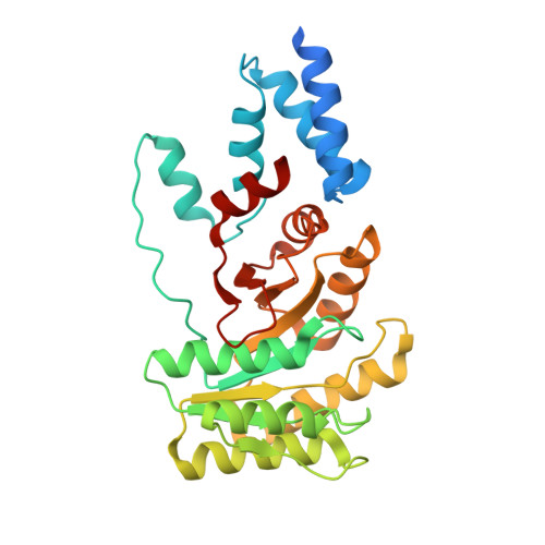
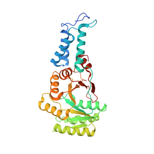
Substrate twinning activates the signal recognition particle and its receptor

Egea, P.F.Shan, S.O.Napetschnig, J.Savage, D.F.Walter, P.Stroud, R.M.

(2004) Nature 427: 215-221

  • DOI: https://doi.org/10.1038/nature02250
  • Primary Citation of Related Structures:  
    1RJ9

  • PubMed Abstract: 

    Signal sequences target proteins for secretion from cells or for integration into cell membranes. As nascent proteins emerge from the ribosome, signal sequences are recognized by the signal recognition particle (SRP), which subsequently associates with its receptor (SR). In this complex, the SRP and SR stimulate each other's GTPase activity, and GTP hydrolysis ensures unidirectional targeting of cargo through a translocation pore in the membrane. To define the mechanism of reciprocal activation, we determined the 1.9 A structure of the complex formed between these two GTPases. The two partners form a quasi-two-fold symmetrical heterodimer. Biochemical analysis supports the importance of the extensive interaction surface. Complex formation aligns the two GTP molecules in a symmetrical, composite active site, and the 3'OH groups are essential for association, reciprocal activation and catalysis. This unique circle of twinned interactions is severed twice on hydrolysis, leading to complex dissociation after cargo delivery.


  • Organizational Affiliation

    Department of Biochemistry and Biophysics, University of California at San Francisco, California 94143-2240, USA. pascal@msg.ucsf.edu


Macromolecules
Find similar proteins by:  (by identity cutoff)  |  3D Structure
Entity ID: 1
MoleculeChains Sequence LengthOrganismDetailsImage
Signal Recognition Protein304Thermus aquaticusMutation(s): 1 
UniProt
Find proteins for P83749 (Thermus aquaticus)
Explore P83749 
Go to UniProtKB:  P83749
Entity Groups  
Sequence Clusters30% Identity50% Identity70% Identity90% Identity95% Identity100% Identity
UniProt GroupP83749
Sequence Annotations
Expand
  • Reference Sequence
Find similar proteins by:  (by identity cutoff)  |  3D Structure
Entity ID: 2
MoleculeChains Sequence LengthOrganismDetailsImage
Signal recognition particle protein300Thermus aquaticusMutation(s): 1 
Gene Names: FFH
UniProt
Find proteins for O07347 (Thermus aquaticus)
Explore O07347 
Go to UniProtKB:  O07347
Entity Groups  
Sequence Clusters30% Identity50% Identity70% Identity90% Identity95% Identity100% Identity
UniProt GroupO07347
Sequence Annotations
Expand
  • Reference Sequence
Experimental Data & Validation

Experimental Data

  • Method: X-RAY DIFFRACTION
  • Resolution: 1.90 Å
  • R-Value Free: 0.239 
  • R-Value Work: 0.206 
  • R-Value Observed: 0.239 
  • Space Group: P 21 21 21
Unit Cell:
Length ( Å )Angle ( ˚ )
a = 75.04α = 90
b = 83.68β = 90
c = 94.02γ = 90
Software Package:
Software NamePurpose
DENZOdata reduction
SCALEPACKdata scaling
AMoREphasing
CNSrefinement

Structure Validation

View Full Validation Report


Ligand Structure Quality Assessment 


Entry History 

Deposition Data

Revision History  (Full details and data files)

  • Version 1.0: 2004-01-27
    Type: Initial release
  • Version 1.1: 2008-04-29
    Changes: Version format compliance
  • Version 1.2: 2011-07-13
    Changes: Version format compliance
  • Version 1.3: 2021-10-27
    Changes: Database references, Derived calculations
  • Version 1.4: 2024-02-14
    Changes: Data collection
  • Version 1.5: 2024-04-03
    Changes: Refinement description