1RIT

Crystal structure of Peanut lectin in complex with meso-tetrasulphonatophenylporphyrin and lactose


Experimental Data Snapshot

  • Method: X-RAY DIFFRACTION
  • Resolution: 2.85 Å
  • R-Value Free: 0.260 
  • R-Value Work: 0.235 
  • R-Value Observed: 0.235 

wwPDB Validation   3D Report Full Report


Ligand Structure Quality Assessment 


This is version 2.1 of the entry. See complete history


Literature

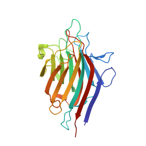
Crystal structures of the PNA-porphyrin complex in the presence and absence of lactose: mapping the conformational changes on lactose binding, interacting surfaces, and supramolecular aggregations.

Goel, M.Damai, R.S.Sethi, D.K.Kaur, K.J.Maiya, B.G.Swamy, M.J.Salunke, D.M.

(2005) Biochemistry 44: 5588-5596

  • DOI: https://doi.org/10.1021/bi047377s
  • Primary Citation of Related Structures:  
    1RIR, 1RIT

  • PubMed Abstract: 

    The extraordinary recognition specificity of lectins for carbohydrate ligands appears to be violated as they also bind to porphyrins and other noncarbohydrate ligands. In this study, crystal structures of meso-tetrasulfonatophenylporphyrin (H(2)TPPS) bound to peanut agglutinin (PNA) in the presence and absence of lactose were determined. The binding of H(2)TPPS with PNA involved 11 molecules of H(2)TPPS in different supramolecular stacking arrangements associated with a tetramer of PNA in the crystals of the PNA-H(2)TPPS binary complex as well as the PNA-H(2)TPPS-lactose ternary complex. The ternary complex involved lactose binding only to two subunits of the PNA tetramer, which did not have porphyrin interacting in the vicinity of the carbohydrate-binding site. Comparison of the two structures highlighted the plasticity of the carbohydrate-binding site expressed in terms of the conformational change in lactose binding. The unusual quaternary structure of PNA, which results in exposed protein-protein interaction sites, might be responsible for the porphyrin binding. The association of porphyrin in diverse oligomeric stacking arrangements observed in the PNA-H(2)TPPS complex suggested the possibility of protein-porphyrin aggregation under abnormal physiological conditions. The structures described here provide a possible native conformation of the carbohydrate-binding site of PNA in the absence of the ligand, highlight mapping of the unsaturated binding surfaces of PNA using porphyrin interactions, indicate new leads toward possible application of this lectin in photodynamic therapy, and exhibit diverse modes of porphyrin-lectin interactions with implications to porphyria, a disease that results from abnormal accumulation of porphyrins.


  • Organizational Affiliation

    National Institute of Immunology, New Delhi 110067, India.


Macromolecules
Find similar proteins by:  (by identity cutoff)  |  3D Structure
Entity ID: 1
MoleculeChains Sequence LengthOrganismDetailsImage
Galactose-binding lectin
A, B, C, D
236Arachis hypogaeaMutation(s): 0 
UniProt
Find proteins for P02872 (Arachis hypogaea)
Explore P02872 
Go to UniProtKB:  P02872
Entity Groups  
Sequence Clusters30% Identity50% Identity70% Identity90% Identity95% Identity100% Identity
UniProt GroupP02872
Sequence Annotations
Expand
  • Reference Sequence
Oligosaccharides

Help

Entity ID: 2
MoleculeChains Length2D Diagram Glycosylation3D Interactions
beta-D-galactopyranose-(1-4)-beta-D-glucopyranose
E, F
2N/A
Glycosylation Resources
GlyTouCan:  G84224TW
GlyCosmos:  G84224TW
GlyGen:  G84224TW
Small Molecules
Ligands 3 Unique
IDChains Name / Formula / InChI Key2D Diagram3D Interactions
SFP
Query on SFP

Download Ideal Coordinates CCD File 
G [auth A]
H [auth A]
K [auth A]
L [auth A]
M [auth B]
G [auth A],
H [auth A],
K [auth A],
L [auth A],
M [auth B],
N [auth B],
O [auth B],
R [auth C],
S [auth C],
V [auth D],
W [auth D]
5,10,15,20-TETRAKIS(4-SULPFONATOPHENYL)-21H,23H-PORPHINE
C44 H34 N4 O12 S4
OVPFWIRROXFHGQ-LWQDQPMZSA-J
MN
Query on MN

Download Ideal Coordinates CCD File 
J [auth A],
Q [auth B],
U [auth C],
Y [auth D]
MANGANESE (II) ION
Mn
WAEMQWOKJMHJLA-UHFFFAOYSA-N
CA
Query on CA

Download Ideal Coordinates CCD File 
I [auth A],
P [auth B],
T [auth C],
X [auth D]
CALCIUM ION
Ca
BHPQYMZQTOCNFJ-UHFFFAOYSA-N
Biologically Interesting Molecules (External Reference) 1 Unique
Experimental Data & Validation

Experimental Data

  • Method: X-RAY DIFFRACTION
  • Resolution: 2.85 Å
  • R-Value Free: 0.260 
  • R-Value Work: 0.235 
  • R-Value Observed: 0.235 
  • Space Group: P 32
Unit Cell:
Length ( Å )Angle ( ˚ )
a = 94.9α = 90
b = 94.9β = 90
c = 144.1γ = 120
Software Package:
Software NamePurpose
DENZOdata reduction
SCALEPACKdata scaling
AMoREphasing
CNSrefinement

Structure Validation

View Full Validation Report


Ligand Structure Quality Assessment 


Entry History 

Deposition Data

Revision History  (Full details and data files)

  • Version 1.0: 2004-12-28
    Type: Initial release
  • Version 1.1: 2008-04-29
    Changes: Version format compliance
  • Version 1.2: 2011-07-13
    Changes: Non-polymer description, Version format compliance
  • Version 2.0: 2020-07-29
    Type: Remediation
    Reason: Carbohydrate remediation
    Changes: Atomic model, Data collection, Derived calculations, Non-polymer description, Structure summary
  • Version 2.1: 2024-02-14
    Changes: Data collection, Database references, Structure summary