Hydrolysis of third-generation cephalosporins by class C beta-lactamases. Structures of a transition state analog of cefotoxamine in wild-type and extended spectrum enzymes.
Nukaga, M., Kumar, S., Nukaga, K., Pratt, R.F., Knox, J.R.(2004) J Biol Chem 279: 9344-9352
- PubMed: 14660590
- DOI: https://doi.org/10.1074/jbc.M312356200
- Primary Citation of Related Structures:
1RGY, 1RGZ - PubMed Abstract:
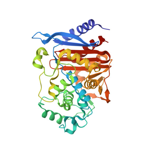
Bacterial resistance to the third-generation cephalosporins is an issue of great concern in current antibiotic therapeutics. An important source of this resistance is from production of extended-spectrum (ES) beta-lactamases by bacteria. The Enterobacter cloacae GC1 enzyme is an example of a class C ES beta-lactamase. Unlike wild-type (WT) forms, such as the E. cloacae P99 and Citrobacter freundii enzymes, the ES GC1 beta-lactamase is able to rapidly hydrolyze third-generation cephalosporins such as cefotaxime and ceftazidime. To understand the basis for this ES activity, m-nitrophenyl 2-(2-aminothiazol-4-yl)-2-[(Z)-methoxyimino]acetylaminomethyl phosphonate has been synthesized and characterized. This phosphonate was designed to generate a transition state analog for turnover of cefotaxime. The crystal structures of complexes of the phosphonate with both ES GC1 and WT C. freundii GN346 beta-lactamases have been determined to high resolution (1.4-1.5 Angstroms). The serine-bound analog of the tetrahedral transition state for deacylation exhibits a very different binding geometry in each enzyme. In the WT beta-lactamase the cefotaxime-like side chain is crowded against the Omega loop and must protrude from the binding site with its methyloxime branch exposed. In the ES enzyme, a mutated Omega loop adopts an alternate conformation allowing the side chain to be much more buried. During the binding and turnover of the cefotaxime substrate by this ES enzyme, it is proposed that ligand-protein contacts and intra-ligand contacts are considerably relieved relative to WT, facilitating positioning and activation of the hydrolytic water molecule. The ES beta-lactamase is thus able to efficiently inactivate third-generation cephalosporins.
Organizational Affiliation:
Department of Molecular and Cell Biology, The University of Connecticut, Storrs, Connecticut 06269-3125, USA.