1RFE

Crystal structure of conserved hypothetical protein Rv2991 from Mycobacterium tuberculosis


Experimental Data Snapshot

  • Method: X-RAY DIFFRACTION
  • Resolution: 2.00 Å
  • R-Value Free: 0.281 
  • R-Value Work: 0.202 
  • R-Value Observed: 0.206 

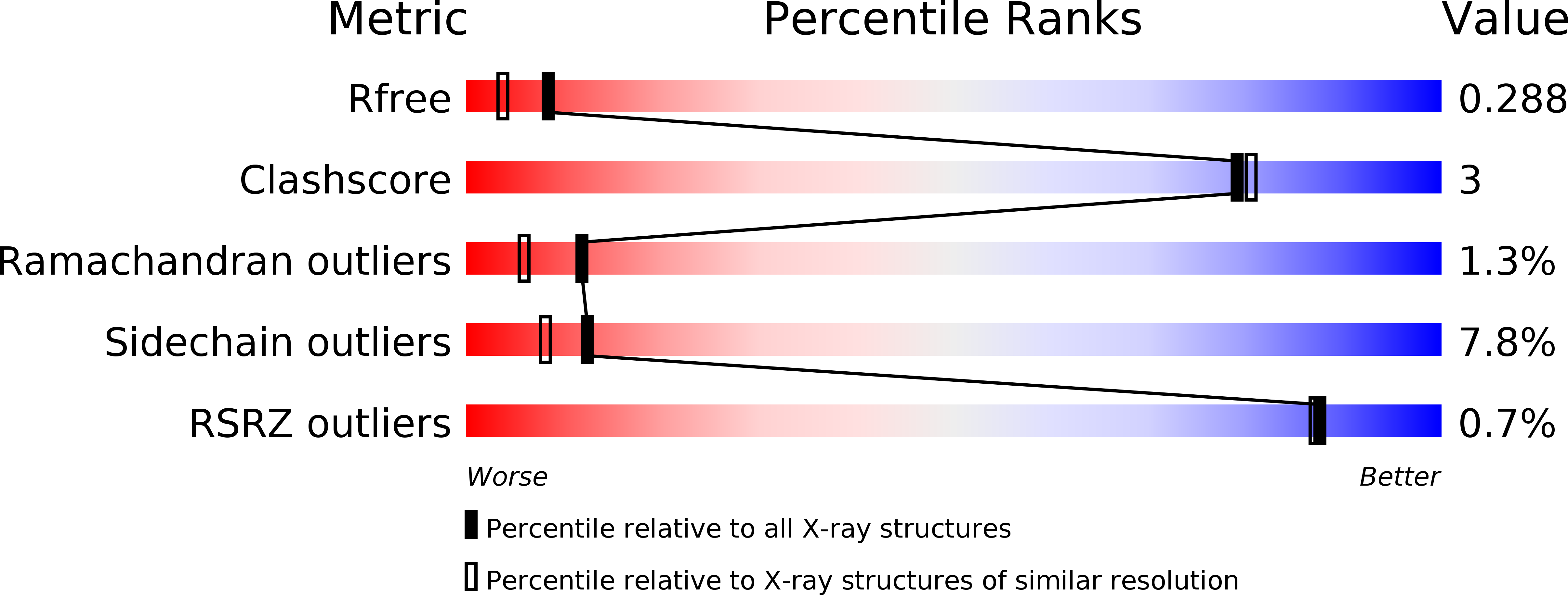
wwPDB Validation   3D Report Full Report


This is version 1.3 of the entry. See complete history


Literature

The crystal structure of Rv2991 from Mycobacterium tuberculosis: An F420binding protein with unknown function.

Benini, S.Haouz, A.Proux, F.Alzari, P.Wilson, K.

(2019) J Struct Biol 

  • DOI: https://doi.org/10.1016/j.jsb.2019.03.006
  • Primary Citation of Related Structures:  
    1RFE

  • PubMed Abstract: 

    The crystal structure of the conserved hypothetical protein Rv2991 from Mycobacterium tuberculosis has been solved by SAD using seleno-methionine substituted protein. The dimeric biological assembly and the sequence and fold conservation are typical of F 420 cofactor binding enzymes. Despite Rv2991 still being of unknown function, sequence and structural comparison with similar proteins enable a role to be proposed for its C-terminal stretch of residues in recognizing and orienting the substrate. In addition, the C-terminus is involved in both protein folding and determining the size of the active site cavity.


  • Organizational Affiliation

    Bioorganic Chemistry and Bio-Crystallography Laboratory (B(2)Cl), Faculty of Science and Technology, Free University of Bolzano, Piazza Università 5, Bolzano 39100, Italy. Electronic address: stefano.benini@unibz.it.


Macromolecules
Find similar proteins by:  (by identity cutoff)  |  3D Structure
Entity ID: 1
MoleculeChains Sequence LengthOrganismDetailsImage
hypothetical protein Rv2991162Mycobacterium tuberculosis H37RvMutation(s): 7 
UniProt
Find proteins for O53240 (Mycobacterium tuberculosis (strain ATCC 25618 / H37Rv))
Explore O53240 
Go to UniProtKB:  O53240
Entity Groups  
Sequence Clusters30% Identity50% Identity70% Identity90% Identity95% Identity100% Identity
UniProt GroupO53240
Sequence Annotations
Expand
  • Reference Sequence
Small Molecules
Modified Residues  2 Unique
IDChains TypeFormula2D DiagramParent
CSX
Query on CSX
A
L-PEPTIDE LINKINGC3 H7 N O3 SCYS
MSE
Query on MSE
A
L-PEPTIDE LINKINGC5 H11 N O2 SeMET
Experimental Data & Validation

Experimental Data

  • Method: X-RAY DIFFRACTION
  • Resolution: 2.00 Å
  • R-Value Free: 0.281 
  • R-Value Work: 0.202 
  • R-Value Observed: 0.206 
  • Space Group: P 43 2 2
Unit Cell:
Length ( Å )Angle ( ˚ )
a = 49.309α = 90
b = 49.309β = 90
c = 132.588γ = 90
Software Package:
Software NamePurpose
REFMACrefinement
DENZOdata reduction
SCALEPACKdata scaling
SHELXDphasing
SHARPphasing
ARP/wARPmodel building

Structure Validation

View Full Validation Report


Entry History 

Deposition Data

Revision History  (Full details and data files)

  • Version 1.0: 2004-12-28
    Type: Initial release
  • Version 1.1: 2008-04-29
    Changes: Version format compliance
  • Version 1.2: 2011-07-13
    Changes: Source and taxonomy, Version format compliance
  • Version 1.3: 2019-04-17
    Changes: Data collection, Database references, Derived calculations