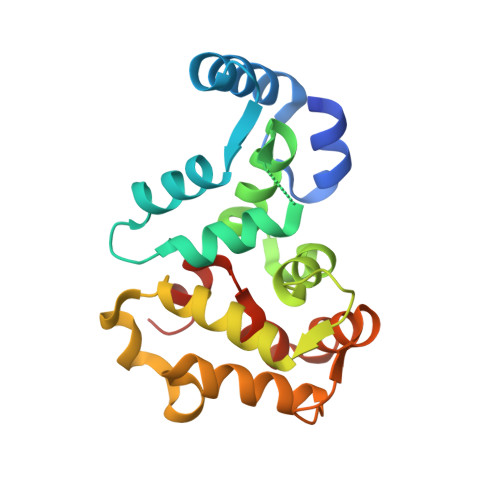
1REC

THREE-DIMENSIONAL STRUCTURE OF RECOVERIN, A CALCIUM SENSOR IN VISION


Experimental Data Snapshot

  • Method: X-RAY DIFFRACTION
  • Resolution: 1.90 Å
  • R-Value Work: 0.191 
  • R-Value Observed: 0.191 

wwPDB Validation   3D Report Full Report


This is version 1.3 of the entry. See complete history


Literature

Three-Dimensional Structure of Recoverin, a Calcium Sensor in Vision

Flaherty, K.M.Zozulya, S.Stryer, L.McKay, D.B.

(1993) Cell 75: 709-716

  • DOI: https://doi.org/10.1016/0092-8674(93)90491-8
  • Primary Citation of Related Structures:  
    1REC

  • PubMed Abstract: 

    Recoverin, a recently discovered member of the EF hand superfamily, serves as a calcium sensor in vision. We report here the crystal structure of recombinant unmyristoylated recoverin at 1.9 A resolution. The four EF hands of the protein are arranged in a compact array that contrasts with the dumbbell shape of calmodulin and troponin C. A calcium ion is bound to EF hand 3, while EF hand 2 can bind samarium but not calcium in this crystal form. The other two EF hands have novel structural features that prevent or impair calcium binding. A concave hydrophobic surface formed by EF hands 1 and 2 may participate in the read out of calcium signals by recoverin and its homologs.


  • Organizational Affiliation

    Beckman Laboratories for Structural Biology, Department of Cell Biology, Stanford University School of Medicine, California 94305.


Macromolecules
Find similar proteins by:  (by identity cutoff)  |  3D Structure
Entity ID: 1
MoleculeChains Sequence LengthOrganismDetailsImage
RECOVERIN201Bos taurusMutation(s): 0 
UniProt
Find proteins for P21457 (Bos taurus)
Explore P21457 
Go to UniProtKB:  P21457
Entity Groups  
Sequence Clusters30% Identity50% Identity70% Identity90% Identity95% Identity100% Identity
UniProt GroupP21457
Sequence Annotations
Expand
  • Reference Sequence
Small Molecules
Ligands 1 Unique
IDChains Name / Formula / InChI Key2D Diagram3D Interactions
CA
Query on CA

Download Ideal Coordinates CCD File 
B [auth A]CALCIUM ION
Ca
BHPQYMZQTOCNFJ-UHFFFAOYSA-N
Experimental Data & Validation

Experimental Data

  • Method: X-RAY DIFFRACTION
  • Resolution: 1.90 Å
  • R-Value Work: 0.191 
  • R-Value Observed: 0.191 
  • Space Group: I 4
Unit Cell:
Length ( Å )Angle ( ˚ )
a = 85.1α = 90
b = 85.1β = 90
c = 60γ = 90
Software Package:
Software NamePurpose
X-PLORmodel building
X-PLORrefinement
X-PLORphasing

Structure Validation

View Full Validation Report


Entry History 

Deposition Data

Revision History  (Full details and data files)

  • Version 1.0: 1994-01-31
    Type: Initial release
  • Version 1.1: 2008-03-24
    Changes: Version format compliance
  • Version 1.2: 2011-07-13
    Changes: Derived calculations, Version format compliance
  • Version 1.3: 2024-02-14
    Changes: Data collection, Database references, Derived calculations, Other