1RBN

THE STRUCTURE OF RIBONUCLEASE A DERIVATIVE II AT 2.1 ANGSTROMS RESOLUTION


Experimental Data Snapshot

  • Method: X-RAY DIFFRACTION
  • Resolution: 2.10 Å
  • R-Value Work: 0.166 
  • R-Value Observed: 0.166 

wwPDB Validation   3D Report Full Report


This is version 1.2 of the entry. See complete history


Literature

Structure of ribonuclease A derivative II at 2.1-A resolution.

Boque, L.Gracia Coll, M.Vilanova, M.Cuchillo, C.M.Fita, I.

(1994) J Biol Chem 269: 19707-19712

  • Primary Citation of Related Structures:  
    1RBN

  • PubMed Abstract: 

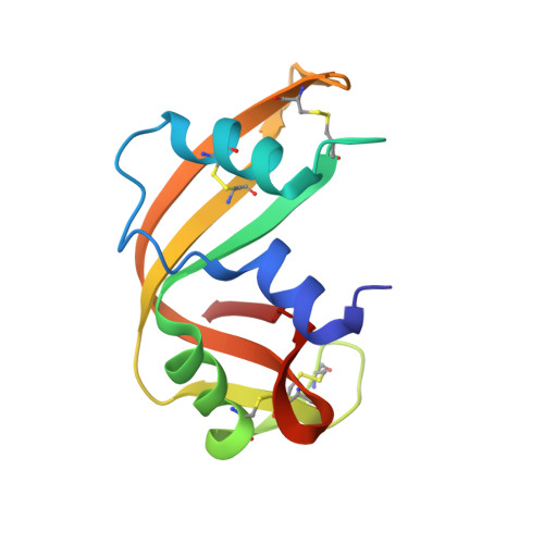
    The crystal structure of bovine pancreatic ribonuclease A derivative II, a covalent derivative obtained by reaction of 6-chloropurine 9-beta-D-ribofuranosyl 5'-monophosphate with the alpha-amino group of Lys-1, has been determined and refined at 2.1-A resolution with an agreement factor R = 0.166 for 6254 reflections in the resolution shell 8.0 to 2.1 A. Crystals are orthorhombic and belong to space group C222(1) with unit cell parameters a = 75.73 A, b = 57.85 A, and c = 53.26 A. This crystal packing had never been reported before for pancreatic ribonuclease nor its complexes. The structure found is in accordance with the location of p2, B3, and R3 subsites at the N-terminal region of the protein and provides an explanation of the catalytic behavior observed for this derivative. In particular, differences in kinetic parameters and in the pKa value of His-119 between derivative II and native ribonuclease A can be interpreted on the basis of the position of the phosphate moiety within the derivative structure. Some uncertainty remains on the nucleotide sugar conformation determined.


  • Organizational Affiliation

    Departament d'Enginyeria Química, Universitat Politècnica de Catalunya, Barcelona, Spain.


Macromolecules
Find similar proteins by:  (by identity cutoff)  |  3D Structure
Entity ID: 1
MoleculeChains Sequence LengthOrganismDetailsImage
RIBONUCLEASE A124Bos taurusMutation(s): 0 
EC: 3.1.27.5
UniProt
Find proteins for P61823 (Bos taurus)
Explore P61823 
Go to UniProtKB:  P61823
Entity Groups  
Sequence Clusters30% Identity50% Identity70% Identity90% Identity95% Identity100% Identity
UniProt GroupP61823
Sequence Annotations
Expand
  • Reference Sequence
Small Molecules
Ligands 1 Unique
IDChains Name / Formula / InChI Key2D Diagram3D Interactions
P5P
Query on P5P

Download Ideal Coordinates CCD File 
B [auth A]PURINE RIBOSIDE-5'-MONOPHOSPHATE
C10 H13 N4 O7 P
MCWDCZIDTUQRHK-FDDDBJFASA-N
Experimental Data & Validation

Experimental Data

  • Method: X-RAY DIFFRACTION
  • Resolution: 2.10 Å
  • R-Value Work: 0.166 
  • R-Value Observed: 0.166 
  • Space Group: C 2 2 21
Unit Cell:
Length ( Å )Angle ( ˚ )
a = 75.73α = 90
b = 57.85β = 90
c = 53.26γ = 90
Software Package:
Software NamePurpose
X-PLORmodel building
X-PLORrefinement
X-PLORphasing

Structure Validation

View Full Validation Report


Entry History 

Deposition Data

Revision History  (Full details and data files)

  • Version 1.0: 1994-12-20
    Type: Initial release
  • Version 1.1: 2008-03-03
    Changes: Version format compliance
  • Version 1.2: 2011-07-13
    Changes: Version format compliance