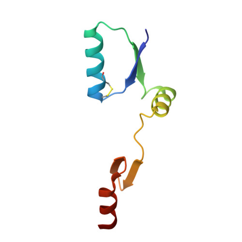
NrdH-redoxin of Corynebacterium ammoniagenes forms a domain-swapped dimer.
Stehr, M., Lindqvist, Y.(2004) Proteins 55: 613-619
- PubMed: 15103625
- DOI: https://doi.org/10.1002/prot.20126
- Primary Citation of Related Structures:
1R7H - PubMed Abstract:
NrdH-redoxins constitute a family of small redox proteins, which contain a conserved CXXC sequence motif, and are characterized by a glutaredoxin-like amino acid sequence but a thioredoxin-like activity profile. Here we report the structure of Corynebacterium ammoniagenes NrdH at 2.7 A resolution, determined by molecular replacement using E. coli NrdH as model. The structure is the first example of a domain-swapped dimer from the thioredoxin family. The domain-swapped structure is formed by an inter-chain two-stranded anti-parallel beta-sheet and is stabilized by electrostatic interactions at the dimer interface. Size exclusion chromatography, and MALDI-ESI experiments revealed however, that the protein exists as a monomer in solution. Similar to E. coli NrdH-redoxin and thioredoxin, C. ammoniagenes NrdH-redoxin has a wide hydrophobic pocket at the surface that could be involved in binding to thioredoxin reductase. However, the loop between alpha2 and beta3, which is complementary to a crevice in the reductase in the thioredoxin-thioredoxin reductase complex, is the hinge for formation of the swapped dimer in C. ammoniagenes NrdH-redoxin. C. ammoniagenes NrdH-redoxin has the highly conserved sequence motif W61-S-G-F-R-P-[DE]67 which is unique to the NrdH-redoxins and which determines the orientation of helix alpha3. An extended hydrogen-bond network, similar to that in E. coli NrdH-redoxin, determines the conformation of the loop formed by the conserved motif.
Organizational Affiliation:
Division of Molecular Structural Biology, Department of Medical Biochemistry and Biophysics, Karolinska Institutet, Stockholm, Sweden.