1R6T

crystal structure of human tryptophanyl-tRNA synthetase


Experimental Data Snapshot

  • Method: X-RAY DIFFRACTION
  • Resolution: 2.10 Å
  • R-Value Free: 0.239 
  • R-Value Work: 0.208 
  • R-Value Observed: 0.208 

wwPDB Validation   3D Report Full Report


Ligand Structure Quality Assessment 


This is version 1.3 of the entry. See complete history


Literature

Crystal structures that suggest late development of genetic code components for differentiating aromatic side chains

Yang, X.-L.Otero, F.J.Skene, R.J.McRee, D.E.Schimmel, P.Ribas De Pouplana, L.

(2003) Proc Natl Acad Sci U S A 100: 15376-15380

  • DOI: https://doi.org/10.1073/pnas.2136794100
  • Primary Citation of Related Structures:  
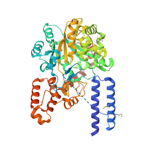
    1Q11, 1R6T

  • PubMed Abstract: 

    Early forms of the genetic code likely generated "statistical" proteins, with similar side chains occupying the same sequence positions at different ratios. In this scenario, groups of related side chains were treated by aminoacyl-tRNA synthetases as a single molecular species until a discrimination mechanism developed that could separate them. The aromatic amino acids tryptophan, tyrosine, and phenylalanine likely constituted one of these groups. A crystal structure of human tryptophanyl-tRNA synthetase was solved at 2.1 A with a tryptophanyl-adenylate bound at the active site. A cocrystal structure of an active fragment of human tyrosyl-tRNA synthetase with its cognate amino acid analog was also solved at 1.6 A. The two structures enabled active site identifications and provided the information for structure-based sequence alignments of approximately 45 orthologs of each enzyme. Two critical positions shared by all tyrosyl-tRNA synthetases and tryptophanyl-tRNA synthetases for amino acid discrimination were identified. The variations at these two positions and phylogenetic analyses based on the structural information suggest that, in contrast to many other amino acids, discrimination of tyrosine from tryptophan occurred late in the development of the genetic code.


  • Organizational Affiliation

    Departments of Molecular Biology and Chemistry, The Scripps Research Institute, BCC-379, 10550 North Torrey Pines Road, La Jolla, CA 92037, USA.


Macromolecules
Find similar proteins by:  (by identity cutoff)  |  3D Structure
Entity ID: 1
MoleculeChains Sequence LengthOrganismDetailsImage
Tryptophanyl-tRNA synthetase
A, B
477Homo sapiensMutation(s): 14 
Gene Names: WARS
EC: 6.1.1.2
UniProt & NIH Common Fund Data Resources
Find proteins for P23381 (Homo sapiens)
Explore P23381 
Go to UniProtKB:  P23381
PHAROS:  P23381
GTEx:  ENSG00000140105 
Entity Groups  
Sequence Clusters30% Identity50% Identity70% Identity90% Identity95% Identity100% Identity
UniProt GroupP23381
Sequence Annotations
Expand
  • Reference Sequence
Small Molecules
Modified Residues  1 Unique
IDChains TypeFormula2D DiagramParent
MSE
Query on MSE
A, B
L-PEPTIDE LINKINGC5 H11 N O2 SeMET
Experimental Data & Validation

Experimental Data

  • Method: X-RAY DIFFRACTION
  • Resolution: 2.10 Å
  • R-Value Free: 0.239 
  • R-Value Work: 0.208 
  • R-Value Observed: 0.208 
  • Space Group: C 1 2 1
Unit Cell:
Length ( Å )Angle ( ˚ )
a = 152.146α = 90
b = 95.713β = 91.62
c = 98.506γ = 90
Software Package:
Software NamePurpose
Blu-Icedata collection
SCALEPACKdata scaling
SOLVEphasing
CNSrefinement

Structure Validation

View Full Validation Report


Ligand Structure Quality Assessment 


Entry History 

Deposition Data

Revision History  (Full details and data files)

  • Version 1.0: 2004-01-06
    Type: Initial release
  • Version 1.1: 2008-04-29
    Changes: Version format compliance
  • Version 1.2: 2011-07-13
    Changes: Non-polymer description, Version format compliance
  • Version 1.3: 2021-10-27
    Changes: Database references, Derived calculations