1R6G

Crystal structure of the thyroid hormone receptor beta ligand binding domain in complex with a beta selective compound


Experimental Data Snapshot

  • Method: X-RAY DIFFRACTION
  • Resolution: 3.00 Å
  • R-Value Free: 0.347 
  • R-Value Work: 0.261 
  • R-Value Observed: 0.265 

wwPDB Validation   3D Report Full Report


Ligand Structure Quality Assessment 


This is version 1.3 of the entry. See complete history


Literature

Thyroid receptor ligands. Part 2: Thyromimetics with improved selectivity for the thyroid hormone receptor beta.

Hangeland, J.J.Doweyko, A.M.Dejneka, T.Friends, T.J.Devasthale, P.Mellstrom, K.Sandberg, J.Grynfarb, M.Sack, J.S.Einspahr, H.Farnegardh, M.Husman, B.Ljunggren, J.Koehler, K.Sheppard, C.Malm, J.Ryono, D.E.

(2004) Bioorg Med Chem Lett 14: 3549-3553

  • DOI: https://doi.org/10.1016/j.bmcl.2004.04.032
  • Primary Citation of Related Structures:  
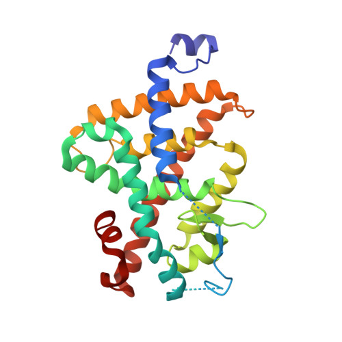
    1R6G

  • PubMed Abstract: 

    A set of thyromimetics having improved selectivity for TR-beta1 were prepared by replacing the 3'-isopropyl group of 2 and 3 with substituents having increased steric bulk. From this limited SAR study, the most potent and selective compounds identified were derived from 2 and contained a 3'-phenyl moiety bearing small hydrophobic groups meta to the biphenyl link. X-ray crystal data of 15c complexed with TR-beta1 LBD shows methionine 442 to be displaced by the bulky R3' phenyl ethyl amide side chain. Movement of this amino acid side chain provides an expanded pocket for the bulky side chain while the ligand-receptor complex retains full agonist activity.


  • Organizational Affiliation

    Pharmaceutical Research Institute, Bristol-Myers Squibb, Princeton, NJ 08543, USA. jon.hangeland@bms.com


Macromolecules
Find similar proteins by:  (by identity cutoff)  |  3D Structure
Entity ID: 1
MoleculeChains Sequence LengthOrganismDetailsImage
Thyroid hormone receptor beta-1259Homo sapiensMutation(s): 0 
Gene Names: THRBNR1A2ERBA2THR1
UniProt & NIH Common Fund Data Resources
Find proteins for P10828 (Homo sapiens)
Explore P10828 
Go to UniProtKB:  P10828
PHAROS:  P10828
GTEx:  ENSG00000151090 
Entity Groups  
Sequence Clusters30% Identity50% Identity70% Identity90% Identity95% Identity100% Identity
UniProt GroupP10828
Sequence Annotations
Expand
  • Reference Sequence
Small Molecules
Ligands 1 Unique
IDChains Name / Formula / InChI Key2D Diagram3D Interactions
442
Query on 442

Download Ideal Coordinates CCD File 
B [auth A]2-[3,5-DIBROMO-4-(4-HYDROXY-3-{HYDROXY[(2-PHENYLETHYL)AMINO]METHYL}PHENOXY)PHENYL]ETHANE-1,1-DIOL
C23 H23 Br2 N O5
KIRQJXNQGZAKGR-UHFFFAOYSA-N
Binding Affinity Annotations 
IDSourceBinding Affinity
442 PDBBind:  1R6G IC50: 0.62 (nM) from 1 assay(s)
Experimental Data & Validation

Experimental Data

  • Method: X-RAY DIFFRACTION
  • Resolution: 3.00 Å
  • R-Value Free: 0.347 
  • R-Value Work: 0.261 
  • R-Value Observed: 0.265 
  • Space Group: P 31 2 1
Unit Cell:
Length ( Å )Angle ( ˚ )
a = 68.556α = 90
b = 68.556β = 90
c = 129.17γ = 120
Software Package:
Software NamePurpose
REFMACrefinement
DENZOdata reduction
SCALEPACKdata scaling
AMoREphasing

Structure Validation

View Full Validation Report


Ligand Structure Quality Assessment 


Entry History 

Revision History  (Full details and data files)

  • Version 1.0: 2005-02-15
    Type: Initial release
  • Version 1.1: 2008-04-29
    Changes: Version format compliance
  • Version 1.2: 2011-07-13
    Changes: Version format compliance
  • Version 1.3: 2024-02-14
    Changes: Data collection, Database references, Derived calculations