Structural role of a buried salt bridge in the 434 repressor DNA-binding domain.
Pervushin, K., Billeter, M., Siegal, G., Wuthrich, K.(1996) J Mol Biol 264: 1002-1012
- PubMed: 9000626
- DOI: https://doi.org/10.1006/jmbi.1996.0692
- Primary Citation of Related Structures:
1R63, 2R63 - PubMed Abstract:
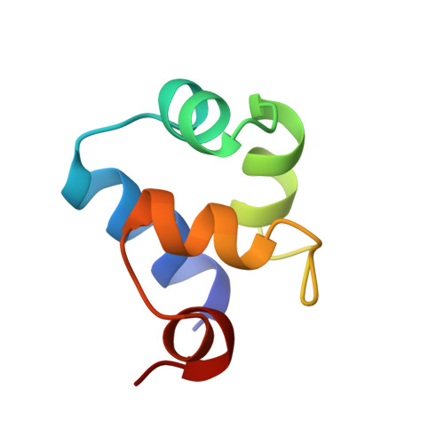
The independently folding 63-residue N-terminal DNA-binding domain of the 434 repressor, 434(1-63), contains a buried Arg10-Glu35 salt bridge. A corresponding salt bridge is found in a variety of prokaryotic and eukaryotic DNA-binding proteins with helix-turn-helix motifs. Here, the NMR solution structures of 434(1-63) and the mutant protein 434[R10M](1-63) were determined to investigate the structural role of this salt bridge. Both proteins contain the same type of global fold, with five alpha-helices and a helix-turn-helix motif formed by the helices II and III. The primary structural difference caused by the Arg10 --> Met mutation is a translation of helix I along its axis relative to the helix II-turn-helix III motif. This limited conformational change is paralleled by a 9 kJ M(-1) decrease of the stability of the folded mutant protein in aqueous solution at pH 4.8. It affects the pKa value of Glu19 as well as the population of a hydrogen bond between the backbone amide proton of Asn16 and the side-chain carboxylate group of Glu19. Using the crystal structure of the 434 repressor dimer complexed with the operator DNA as a basis, model building of the DNA complex with the NMR structure of 434[R10M](1-63) shows that Asn16, which is located on the protein surface, makes direct contact with the DNA and indicates that the point mutation Arg10 --> Met should also lead to modifications of the protein-protein contacts in the complex.
Organizational Affiliation:
Institut für Molekularbiologie und Biophysik, Eidgenössische Technische, Hochschule-Hönggerberg, Zürich, Switzerland.