1R50

Bacillus subtilis lipase A with covalently bound Sc-IPG-phosphonate-inhibitor


Experimental Data Snapshot

  • Method: X-RAY DIFFRACTION
  • Resolution: 1.45 Å
  • R-Value Free: 0.216 
  • R-Value Work: 0.196 

wwPDB Validation   3D Report Full Report


Ligand Structure Quality Assessment 


This is version 1.3 of the entry. See complete history


Literature

Directed Evolution of Bacillus subtilis Lipase A by Use of Enantiomeric Phosphonate Inhibitors: Crystal Structures and Phage Display Selection

Droege, M.J.Boersma, Y.L.van Pouderoyen, G.Vrenken, T.E.Rueggeberg, C.J.Reetz, M.T.Dijkstra, B.W.Quax, W.J.

(2005) Chembiochem 7: 149-157

  • DOI: https://doi.org/10.1002/cbic.200500308
  • Primary Citation of Related Structures:  
    1R4Z, 1R50

  • PubMed Abstract: 

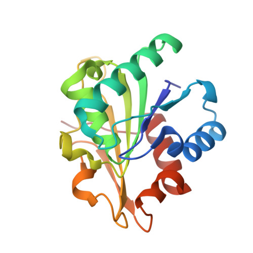
    Phage display can be used as a protein-engineering tool for the selection of proteins with desirable binding properties from a library of mutants. Here we describe the application of this method for the directed evolution of Bacillus subtilis lipase A, an enzyme that has important properties for the preparation of the pharmaceutically relevant chiral compound 1,2-O-isopropylidene-sn-glycerol (IPG). PCR mutagenesis with spiked oligonucleotides was employed for saturation mutagenesis of a stretch of amino acids near the active site. After expression of these mutants on bacteriophages, dual selection with (S)-(+)- and (R)-(-)-IPG stereoisomers covalently coupled to enantiomeric phosphonate suicide inhibitors (SIRAN Sc and Rc inhibitors, respectively) was used for the isolation of variants with inverted enantioselectivity. The mutants were further characterised by determination of their Michaelis-Menten parameters. The 3D structures of the Sc and Rc inhibitor-lipase complexes were determined and provided structural insight into the mechanism of enantioselectivity of the enzyme. In conclusion, we have used phage display as a fast and reproducible method for the selection of Bacillus lipase A mutant enzymes with inverted enantioselectivity.


  • Organizational Affiliation

    Dept. of Pharmaceutical Biology, University of Groningen, A. Deusinglaan 1, 9713 AV Groningen, The Netherlands.


Macromolecules
Find similar proteins by:  (by identity cutoff)  |  3D Structure
Entity ID: 1
MoleculeChains Sequence LengthOrganismDetailsImage
Lipase
A, B
181Bacillus subtilisMutation(s): 0 
Gene Names: lipA
EC: 3.1.1.3
UniProt
Find proteins for P37957 (Bacillus subtilis (strain 168))
Explore P37957 
Go to UniProtKB:  P37957
Entity Groups  
Sequence Clusters30% Identity50% Identity70% Identity90% Identity95% Identity100% Identity
UniProt GroupP37957
Sequence Annotations
Expand
  • Reference Sequence
Small Molecules
Ligands 1 Unique
IDChains Name / Formula / InChI Key2D Diagram3D Interactions
SIL
Query on SIL

Download Ideal Coordinates CCD File 
C [auth A],
D [auth B]
[(4S)-2,2-DIMETHYL-1,3-DIOXOLAN-4-YL]METHYL HYDROGEN HEX-5-ENYLPHOSPHONATE
C12 H23 O5 P
BUTLRPVAJSANIT-LLVKDONJSA-N
Experimental Data & Validation

Experimental Data

  • Method: X-RAY DIFFRACTION
  • Resolution: 1.45 Å
  • R-Value Free: 0.216 
  • R-Value Work: 0.196 
  • Space Group: P 21 21 21
Unit Cell:
Length ( Å )Angle ( ˚ )
a = 39.594α = 90
b = 83.591β = 90
c = 95.082γ = 90
Software Package:
Software NamePurpose
DENZOdata reduction
TRUNCATEdata reduction
CNSrefinement
CCP4data scaling
CNSphasing

Structure Validation

View Full Validation Report


Ligand Structure Quality Assessment 


Entry History 

Deposition Data

Revision History  (Full details and data files)

  • Version 1.0: 2004-10-19
    Type: Initial release
  • Version 1.1: 2008-04-29
    Changes: Version format compliance
  • Version 1.2: 2011-07-13
    Changes: Version format compliance
  • Version 1.3: 2024-04-03
    Changes: Data collection, Database references, Derived calculations, Refinement description, Structure summary