1R2J

FkbI for Biosynthesis of Methoxymalonyl Extender Unit of Fk520 Polyketide Immunosuppresant


Experimental Data Snapshot

  • Method: X-RAY DIFFRACTION
  • Resolution: 2.10 Å
  • R-Value Free: 0.263 
  • R-Value Work: 0.247 
  • R-Value Observed: 0.247 

wwPDB Validation   3D Report Full Report


Ligand Structure Quality Assessment 


This is version 1.6 of the entry. See complete history


Literature

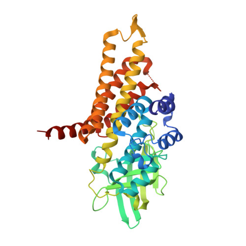
Crystal Structure of an Acyl-ACP Dehydrogenase from the FK520 Polyketide Biosynthetic Pathway: Insights into Extender Unit Biosynthesis

Watanabe, K.Khosla, C.Stroud, R.M.Tsai, S.-C.

(2003) J Mol Biol 334: 435-444

  • DOI: https://doi.org/10.1016/j.jmb.2003.10.021
  • Primary Citation of Related Structures:  
    1R2J

  • PubMed Abstract: 

    Polyketide synthases (PKSs) synthesize the polyketide cores of pharmacologically important natural products such as the immunosuppressants FK520 and FK506. Understanding polyketide biosynthesis at atomic resolution could present new opportunities for chemo-enzymatic synthesis of complex molecules. The crystal structure of FkbI, an enzyme involved in the biosynthesis of the methoxymalonyl extender unit of FK520, was solved to 2.1A with an R(crys) of 24.4%. FkbI has a similar fold to acyl-CoA dehydrogenases. Notwithstanding this similarity, the surface and substrate-binding site of FkbI reveal key differences from other acyl-CoA dehydrogenases, suggesting that FkbI may recognize an acyl-ACP substrate rather than an acyl-CoA substrate. This structural observation coincided the genetic experiment done by Carroll et al. J. Am. Chem. Soc., 124 (2002) 4176. Although an in vitro assay for FkbI remains elusive, the structural basis for the substrate specificity of FkbI is analyzed by a combination of sequence comparison, docking simulations and structural analysis. A biochemical mechanism for the role of FkbI in the biosynthesis of methoxymalonyl-ACP is proposed.


  • Organizational Affiliation

    Department of Chemical Engineering, Stanford University, Stanford, CA 94305-5025, USA.


Macromolecules
Find similar proteins by:  (by identity cutoff)  |  3D Structure
Entity ID: 1
MoleculeChains Sequence LengthOrganismDetailsImage
protein FkbI366Streptomyces hygroscopicusMutation(s): 0 
UniProt
Find proteins for Q9KIE5 (Streptomyces hygroscopicus subsp. ascomyceticus)
Explore Q9KIE5 
Go to UniProtKB:  Q9KIE5
Entity Groups  
Sequence Clusters30% Identity50% Identity70% Identity90% Identity95% Identity100% Identity
UniProt GroupQ9KIE5
Sequence Annotations
Expand
  • Reference Sequence
Small Molecules
Ligands 1 Unique
IDChains Name / Formula / InChI Key2D Diagram3D Interactions
FAD
Query on FAD

Download Ideal Coordinates CCD File 
B [auth A]FLAVIN-ADENINE DINUCLEOTIDE
C27 H33 N9 O15 P2
VWWQXMAJTJZDQX-UYBVJOGSSA-N
Experimental Data & Validation

Experimental Data

  • Method: X-RAY DIFFRACTION
  • Resolution: 2.10 Å
  • R-Value Free: 0.263 
  • R-Value Work: 0.247 
  • R-Value Observed: 0.247 
  • Space Group: I 41
Unit Cell:
Length ( Å )Angle ( ˚ )
a = 116.55α = 90
b = 116.55β = 90
c = 126.16γ = 90
Software Package:
Software NamePurpose
DENZOdata reduction
SCALEPACKdata scaling
CNSrefinement
CNSphasing

Structure Validation

View Full Validation Report


Ligand Structure Quality Assessment 


Entry History 

Deposition Data

Revision History  (Full details and data files)

  • Version 1.0: 2003-11-25
    Type: Initial release
  • Version 1.1: 2008-04-29
    Changes: Version format compliance
  • Version 1.2: 2011-07-13
    Changes: Version format compliance
  • Version 1.3: 2018-01-31
    Changes: Experimental preparation
  • Version 1.4: 2018-04-04
    Changes: Data collection
  • Version 1.5: 2018-04-11
    Changes: Data collection
  • Version 1.6: 2024-02-14
    Changes: Data collection, Database references, Derived calculations