A metal-ligand-mediated intersubunit allosteric switch in related SmtB/ArsR zinc sensor proteins.
Eicken, C., Pennella, M.A., Chen, X., Koshlap, K.M., VanZile, M.L., Sacchettini, J.C., Giedroc, D.P.(2003) J Mol Biol 333: 683-695
- PubMed: 14568530
- DOI: https://doi.org/10.1016/j.jmb.2003.09.007
- Primary Citation of Related Structures:
1R1T, 1R1U, 1R1V, 1R22, 1R23 - PubMed Abstract:
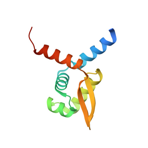
The origin of metal ion selectivity by members of the SmtB/ArsR family of bacterial metal-sensing transcriptional repressors and the mechanism of negative allosteric regulation of DNA binding is poorly understood. Here, we report that two homologous zinc sensors, Staphylococcus aureus CzrA and cyanobacterial SmtB, are "winged" helix homodimeric DNA-binding proteins that bind Zn(II) to a pair of tetrahedral, interhelical binding sites, with two ligands derived from the alpha5 helix of one subunit, Asp84 O(delta1) (Asp104 in SmtB), His86 N(delta1) (His106), and two derived from the alpha5 helix of the other, His97' N(delta1) (His117') and His100' N(epsilon2) (Glu120'). Formation of the metal chelate drives a quaternary structural switch mediated by an intersubunit hydrogen-binding network that originates with the non-liganding N(epsilon2) face of His97 in CzrA (His117 in SmtB) that stabilizes a low-affinity, DNA-binding conformation. The structure of the Zn(1) SmtB homodimer shows that both metal-binding sites of the dimer must be occupied for the quaternary structural switch to occur. Thus, a critical zinc-ligating histidine residue obligatorily couples formation of the metal-sensing coordination chelate to changes in the conformation and dynamics of the putative DNA-binding helices.
Organizational Affiliation:
Department of Biochemistry and Biophysics, Center for Structural Biology, Texas A&M University, College Station, TX 77843-2128, USA.