1QZM

alpha-domain of ATPase


Experimental Data Snapshot

  • Method: X-RAY DIFFRACTION
  • Resolution: 1.90 Å
  • R-Value Free: 0.282 
  • R-Value Work: 0.179 
  • R-Value Observed: 0.184 

wwPDB Validation   3D Report Full Report


This is version 1.4 of the entry. See complete history


Literature

Crystal structure of the AAA+ alpha domain of E. coli Lon protease at 1.9A resolution.

Botos, I.Melnikov, E.E.Cherry, S.Khalatova, A.G.Rasulova, F.S.Tropea, J.E.Maurizi, M.R.Rotanova, T.V.Gustchina, A.Wlodawer, A.

(null) J Struct Biol 146: 113-122

  • DOI: https://doi.org/10.1016/j.jsb.2003.09.003
  • Primary Citation of Related Structures:  
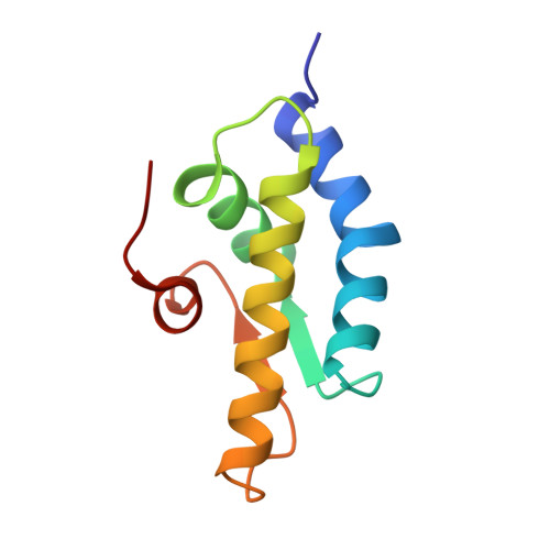
    1QZM

  • PubMed Abstract: 

    The crystal structure of the small, mostly helical alpha domain of the AAA+ module of the Escherichia coli ATP-dependent protease Lon has been solved by single isomorphous replacement combined with anomalous scattering and refined at 1.9A resolution to a crystallographic R factor of 17.9%. This domain, comprising residues 491-584, was obtained by chymotrypsin digestion of the recombinant full-length protease. The alpha domain of Lon contains four alpha helices and two parallel strands and resembles similar domains found in a variety of ATPases and helicases, including the oligomeric proteases HslVU and ClpAP. The highly conserved "sensor-2" Arg residue is located at the beginning of the third helix. Detailed comparison with the structures of 11 similar domains established the putative location of the nucleotide-binding site in this first fragment of Lon for which a crystal structure has become available.


  • Organizational Affiliation

    Macromolecular Crystallography Laboratory, National Cancer Institute at Frederick, MCL Bldg. 536, Rm. 5, Frederick, MD 21702-1201, USA.


Macromolecules
Find similar proteins by:  (by identity cutoff)  |  3D Structure
Entity ID: 1
MoleculeChains Sequence LengthOrganismDetailsImage
ATP-dependent protease La94Escherichia coliMutation(s): 0 
Gene Names: LON
EC: 3.4.21.53
UniProt
Find proteins for P0A9M0 (Escherichia coli (strain K12))
Explore P0A9M0 
Go to UniProtKB:  P0A9M0
Entity Groups  
Sequence Clusters30% Identity50% Identity70% Identity90% Identity95% Identity100% Identity
UniProt GroupP0A9M0
Sequence Annotations
Expand
  • Reference Sequence
Experimental Data & Validation

Experimental Data

  • Method: X-RAY DIFFRACTION
  • Resolution: 1.90 Å
  • R-Value Free: 0.282 
  • R-Value Work: 0.179 
  • R-Value Observed: 0.184 
  • Space Group: P 43 21 2
Unit Cell:
Length ( Å )Angle ( ˚ )
a = 46.43α = 90
b = 46.43β = 90
c = 85.2γ = 90
Software Package:
Software NamePurpose
SHELXmodel building
SHELXL-97refinement
MAR345data collection
SCALEPACKdata scaling
SHARPphasing
CNSrefinement
SHELXLrefinement

Structure Validation

View Full Validation Report


Entry History 

Deposition Data

Revision History  (Full details and data files)

  • Version 1.0: 2004-05-04
    Type: Initial release
  • Version 1.1: 2007-11-14
    Changes: Version format compliance
  • Version 1.2: 2011-07-13
    Changes: Version format compliance
  • Version 1.3: 2017-10-11
    Changes: Refinement description
  • Version 1.4: 2024-02-14
    Changes: Data collection, Database references