1QYN

Crystal Structure of SecB from Escherichia coli


Experimental Data Snapshot

  • Method: X-RAY DIFFRACTION
  • Resolution: 2.35 Å
  • R-Value Free: 0.271 
  • R-Value Work: 0.234 
  • R-Value Observed: 0.234 

wwPDB Validation   3D Report Full Report


This is version 1.4 of the entry. See complete history


Literature

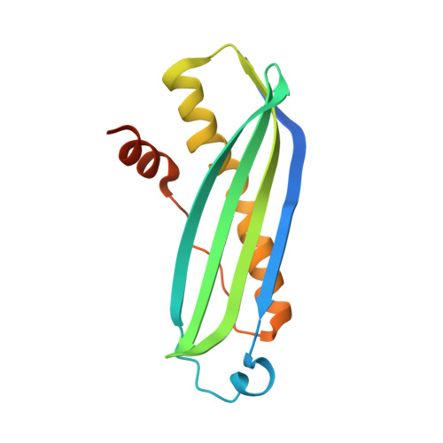
Crystal structure of SecB from Escherichia coli

Dekker, C.de Kruijff, B.Gros, P.

(2003) J Struct Biol 144: 313-319

  • DOI: https://doi.org/10.1016/j.jsb.2003.09.012
  • Primary Citation of Related Structures:  
    1QYN

  • PubMed Abstract: 

    The chaperone SecB from Escherichia coli is primarily involved in passing precursor proteins into the Sec system via specific interactions with SecA. The crystal structure of SecB from E. coli has been solved to 2.35 A resolution. The structure shows flexibility in the crossover loop and the helix-connecting loop, regions that have been implicated to be part of the SecB substrate-binding site. Moreover conformational variability of Trp36 is observed as well as different loop conformations for the different monomers. Based on this, we speculate that SecB can regulate the access or extent of its hydrophobic substrate-binding site, by modulating the conformation of the crossover loop and the helix-connecting loop. The structure also clearly explains why the tetrameric equilibrium is shifted towards the dimeric state in the mutant SecBCys76Tyr. The buried cysteine residue is crucial for tight packing, and mutations are likely to disrupt the tetramer formation but not the dimer formation.


  • Organizational Affiliation

    National Institute for Medical Research, The Ridgeway, Mill Hill, London, England, NW7 1AA, UK. cdekker@mimr.mrc.ac.uk


Macromolecules
Find similar proteins by:  (by identity cutoff)  |  3D Structure
Entity ID: 1
MoleculeChains Sequence LengthOrganismDetailsImage
Protein-export protein secB
A, B, C, D
153Escherichia coliMutation(s): 0 
Gene Names: SECB
UniProt
Find proteins for P0AG86 (Escherichia coli (strain K12))
Explore P0AG86 
Go to UniProtKB:  P0AG86
Entity Groups  
Sequence Clusters30% Identity50% Identity70% Identity90% Identity95% Identity100% Identity
UniProt GroupP0AG86
Sequence Annotations
Expand
  • Reference Sequence
Experimental Data & Validation

Experimental Data

  • Method: X-RAY DIFFRACTION
  • Resolution: 2.35 Å
  • R-Value Free: 0.271 
  • R-Value Work: 0.234 
  • R-Value Observed: 0.234 
  • Space Group: P 43 21 2
Unit Cell:
Length ( Å )Angle ( ˚ )
a = 84.005α = 90
b = 84.005β = 90
c = 204.9γ = 90
Software Package:
Software NamePurpose
DENZOdata reduction
SCALEPACKdata scaling
AMoREphasing
CNSrefinement

Structure Validation

View Full Validation Report


Entry History 

Deposition Data

Revision History  (Full details and data files)

  • Version 1.0: 2003-12-09
    Type: Initial release
  • Version 1.1: 2008-04-29
    Changes: Version format compliance
  • Version 1.2: 2011-07-13
    Changes: Version format compliance
  • Version 1.3: 2017-10-11
    Changes: Refinement description
  • Version 1.4: 2023-08-23
    Changes: Data collection, Database references, Refinement description