1QXY

Crystal structure of S. aureus methionine aminopeptidase in complex with a ketoheterocycle 618


Experimental Data Snapshot

  • Method: X-RAY DIFFRACTION
  • Resolution: 1.04 Å
  • R-Value Free: 0.165 
  • R-Value Work: 0.142 
  • R-Value Observed: 0.144 

wwPDB Validation   3D Report Full Report


Ligand Structure Quality Assessment 


This is version 1.3 of the entry. See complete history


Literature

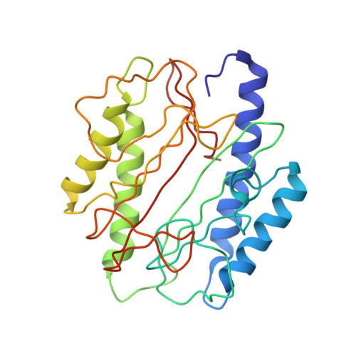
Crystal structures of staphylococcusaureus methionine aminopeptidase complexed with keto heterocycle and aminoketone inhibitors reveal the formation of a tetrahedral intermediate.

Douangamath, A.Dale, G.E.D'Arcy, A.Almstetter, M.Eckl, R.Frutos-Hoener, A.Henkel, B.Illgen, K.Nerdinger, S.Schulz, H.MacSweeney, A.Thormann, M.Treml, A.Pierau, S.Wadman, S.Oefner, C.

(2004) J Med Chem 47: 1325-1328

  • DOI: https://doi.org/10.1021/jm034188j
  • Primary Citation of Related Structures:  
    1QXW, 1QXY, 1QXZ

  • PubMed Abstract: 

    High-resolution crystal structures of Staphylococcus aureus methionine aminopeptidase I in complex with various keto heterocycles and aminoketones were determined, and the intermolecular ligand interactions with the enzyme are reported. The compounds are effective inhibitors of the S. aureus enzyme because of the formation of an uncleavable tetrahedral intermediate upon binding. The electron densities unequivocally show the enzyme-catalyzed transition-state analogue mimicking that for amide bond hydrolysis of substrates.


  • Organizational Affiliation

    Morphochem AG, WRO-1055/388, Schwarzwaldallee 215, CH-4058 Basel, Switzerland.


Macromolecules
Find similar proteins by:  (by identity cutoff)  |  3D Structure
Entity ID: 1
MoleculeChains Sequence LengthOrganismDetailsImage
methionyl aminopeptidase252Staphylococcus aureusMutation(s): 0 
EC: 3.4.11.18
UniProt
Find proteins for P0A078 (Staphylococcus aureus (strain Mu50 / ATCC 700699))
Explore P0A078 
Go to UniProtKB:  P0A078
Entity Groups  
Sequence Clusters30% Identity50% Identity70% Identity90% Identity95% Identity100% Identity
UniProt GroupP0A078
Sequence Annotations
Expand
  • Reference Sequence
Small Molecules
Binding Affinity Annotations 
IDSourceBinding Affinity
M2C Binding MOAD:  1QXY IC50: 1.60e+4 (nM) from 1 assay(s)
PDBBind:  1QXY IC50: 1.60e+4 (nM) from 1 assay(s)
Experimental Data & Validation

Experimental Data

  • Method: X-RAY DIFFRACTION
  • Resolution: 1.04 Å
  • R-Value Free: 0.165 
  • R-Value Work: 0.142 
  • R-Value Observed: 0.144 
  • Space Group: P 1 21 1
Unit Cell:
Length ( Å )Angle ( ˚ )
a = 40.97α = 90
b = 76.69β = 104.5
c = 41.66γ = 90
Software Package:
Software NamePurpose
REFMACrefinement
XDSdata reduction
CCP4data scaling
AMoREphasing

Structure Validation

View Full Validation Report


Ligand Structure Quality Assessment 


Entry History 

Deposition Data

Revision History  (Full details and data files)

  • Version 1.0: 2004-03-16
    Type: Initial release
  • Version 1.1: 2008-04-29
    Changes: Version format compliance
  • Version 1.2: 2011-07-13
    Changes: Version format compliance
  • Version 1.3: 2024-02-14
    Changes: Data collection, Database references, Derived calculations