1QWI

Crystal Structure of E. coli OsmC


Experimental Data Snapshot

  • Method: X-RAY DIFFRACTION
  • Resolution: 1.80 Å
  • R-Value Free: 0.239 
  • R-Value Work: 0.222 

wwPDB Validation   3D Report Full Report


This is version 1.2 of the entry. See complete history


Literature

Structural and functional features of the Escherichia coli hydroperoxide resistance protein OsmC

Lesniak, J.Barton, W.A.Nikolov, D.B.

(2003) Protein Sci 12: 2838-2843

  • DOI: https://doi.org/10.1110/ps.03375603
  • Primary Citation of Related Structures:  
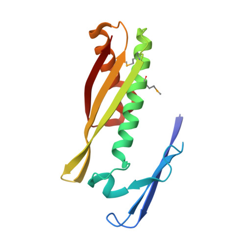
    1QWI

  • PubMed Abstract: 

    The osmotically inducible protein OsmC, like its better-characterized homolog, the organic hydroperoxide protein Ohr, is involved in defense against oxidative stress caused by exposure to organic hydroperoxides. The crystal structure of Escherichia coli OsmC reported here reveals that the protein is a tightly folded domain-swapped dimer with two active sites located at the monomer interface on opposite sides of the molecule. We demonstrate that OsmC preferentially metabolizes organic hydroperoxides over inorganic hydrogen peroxide. On the basis of structural and enzymatic similarities, we propose that the OsmC catalytic mechanism is analogous to that of the Ohr proteins and of the structurally unrelated peroxiredoxins, directly using highly reactive cysteine thiol groups to elicit hydroperoxide reduction.


  • Organizational Affiliation

    Joan and Sanford I. Weill Graduate School of Medical Sciences of Cornell University, New York, New York 10021, USA.


Macromolecules
Find similar proteins by:  (by identity cutoff)  |  3D Structure
Entity ID: 1
MoleculeChains Sequence LengthOrganismDetailsImage
osmotically inducible protein
A, B, C, D
143Escherichia coliMutation(s): 3 
Gene Names: OsmC
UniProt
Find proteins for P0C0L2 (Escherichia coli (strain K12))
Explore P0C0L2 
Go to UniProtKB:  P0C0L2
Entity Groups  
Sequence Clusters30% Identity50% Identity70% Identity90% Identity95% Identity100% Identity
UniProt GroupP0C0L2
Sequence Annotations
Expand
  • Reference Sequence
Small Molecules
Modified Residues  1 Unique
IDChains TypeFormula2D DiagramParent
MSE
Query on MSE
A, B, C, D
L-PEPTIDE LINKINGC5 H11 N O2 SeMET
Experimental Data & Validation

Experimental Data

  • Method: X-RAY DIFFRACTION
  • Resolution: 1.80 Å
  • R-Value Free: 0.239 
  • R-Value Work: 0.222 
  • Space Group: P 1 21 1
Unit Cell:
Length ( Å )Angle ( ˚ )
a = 65.747α = 90
b = 64.229β = 92.62
c = 67.585γ = 90
Software Package:
Software NamePurpose
DENZOdata reduction
SCALEPACKdata scaling
SOLVEphasing
CNSrefinement

Structure Validation

View Full Validation Report


Entry History 

Deposition Data

Revision History  (Full details and data files)

  • Version 1.0: 2003-12-16
    Type: Initial release
  • Version 1.1: 2008-04-29
    Changes: Version format compliance
  • Version 1.2: 2011-07-13
    Changes: Version format compliance