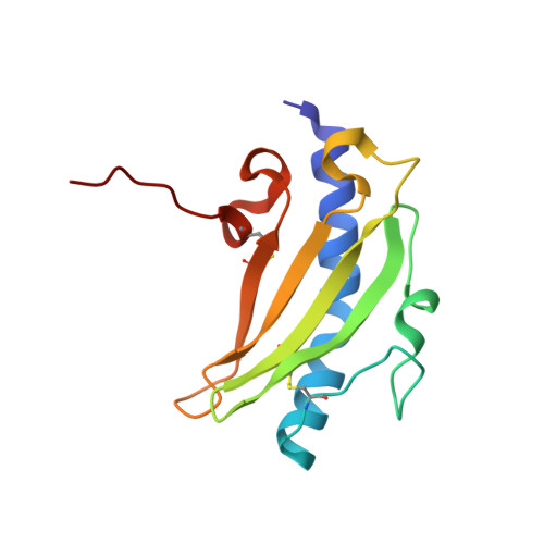
Crystallographic Analysis of the Pseudomonas aeruginosa Strain K122-4 Monomeric Pilin Reveals a Conserved Receptor-Binding Architecture
Audette, G.F., Irvin, R.T., Hazes, B.(2004) Biochemistry 43: 11427-11435
- PubMed: 15350129
- DOI: https://doi.org/10.1021/bi048957s
- Primary Citation of Related Structures:
1QVE, 1RG0 - PubMed Abstract:
Adherence of pathogens to host cells is critical for the initiation of infection and is thus an attractive target for anti-infective therapeutics and vaccines. In the opportunistic human pathogen Pseudomonas aeruginosa, host-cell adherence is achieved predominantly by type IV pili. Analysis of several clinical strains of P. aeruginosa reveals poor sequence conservation between pilin genes, including the residues in the receptor-binding site. Interestingly, the receptor-binding sites appear to retain a conserved surface epitope because all Pseudomonas type IV pili recognize the same receptor on the host cell and cross-reactive antibodies specific for the receptor-binding site exist. Here, we present the crystallographic analysis of two crystal forms of truncated pilin from P. aeruginosa strain K122-4 (DeltaK122-4) at 1.54 and 1.8 A resolution, respectively. The DeltaK122-4 structure is compared to other crystallographically determined type IV pilin structures and an NMR structure of DeltaK122-4 pilin. A comparison with the structure of the highly divergent P. aeruginosa strain K (DeltaPAK) pilin indicates that the receptor-binding loop in both pilins forms a shallow depression with a surface that is formed by main-chain atoms. Conservation of this putative binding site is independent of the sequence as long as the main-chain conformation is conserved and could therefore explain the shared receptor specificity and antibody cross reactivity of highly divergent Pseudomonas type IV pilins.
Organizational Affiliation:
Department of Medical Microbiology and Immunology, University of Alberta, Edmonton, Alberta, Canada, T6G 2H7.