1QUS

1.7 A RESOLUTION STRUCTURE OF THE SOLUBLE LYTIC TRANSGLYCOSYLASE SLT35 FROM ESCHERICHIA COLI


Experimental Data Snapshot

  • Method: X-RAY DIFFRACTION
  • Resolution: 1.70 Å
  • R-Value Free: 0.207 
  • R-Value Work: 0.176 
  • R-Value Observed: 0.178 

wwPDB Validation   3D Report Full Report


Ligand Structure Quality Assessment 


This is version 1.4 of the entry. See complete history


Literature

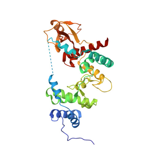
Crystal structure of Escherichia coli lytic transglycosylase Slt35 reveals a lysozyme-like catalytic domain with an EF-hand.

van Asselt, E.J.Dijkstra, A.J.Kalk, K.H.Takacs, B.Keck, W.Dijkstra, B.W.

(1999) Structure 7: 1167-1180

  • DOI: https://doi.org/10.1016/s0969-2126(00)80051-9
  • Primary Citation of Related Structures:  
    1QUS

  • PubMed Abstract: 

    Lytic transglycosylases are bacterial muramidases that catalyse the cleavage of the beta- 1,4-glycosidic bond between N-acetylmuramic acid (MurNAc) and N-acetylglucosamine (GlcNAc) in peptidoglycan with concomitant formation of a 1,6-anhydrobond in the MurNAc residue. These muramidases play an important role in the metabolism of the bacterial cell wall and might therefore be potential targets for the rational design of antibacterial drugs. One of the lytic transglycosylases is Slt35, a naturally occurring soluble fragment of the outer membrane bound lytic transglycosylase B (MltB) from Escherichia coli.


  • Organizational Affiliation

    BIOSON Research Institute, Laboratory of Biophysical Chemistry Groningen University, Nijenborgh 4, 9747 AG, Groningen, The Netherlands.


Macromolecules
Find similar proteins by:  (by identity cutoff)  |  3D Structure
Entity ID: 1
MoleculeChains Sequence LengthOrganismDetailsImage
LYTIC MUREIN TRANSGLYCOSYLASE B322Escherichia coliMutation(s): 2 
EC: 3.2.1
UniProt
Find proteins for P41052 (Escherichia coli (strain K12))
Explore P41052 
Go to UniProtKB:  P41052
Entity Groups  
Sequence Clusters30% Identity50% Identity70% Identity90% Identity95% Identity100% Identity
UniProt GroupP41052
Sequence Annotations
Expand
  • Reference Sequence
Experimental Data & Validation

Experimental Data

  • Method: X-RAY DIFFRACTION
  • Resolution: 1.70 Å
  • R-Value Free: 0.207 
  • R-Value Work: 0.176 
  • R-Value Observed: 0.178 
  • Space Group: P 21 21 21
Unit Cell:
Length ( Å )Angle ( ˚ )
a = 58.329α = 90
b = 67.883β = 90
c = 98.853γ = 90
Software Package:
Software NamePurpose
DMmodel building
WARPmodel building
X-PLORrefinement
DENZOdata reduction
SCALEPACKdata scaling
DMphasing
ARP/wARPmodel building

Structure Validation

View Full Validation Report


Ligand Structure Quality Assessment 


Entry History 

Deposition Data

Revision History  (Full details and data files)

  • Version 1.0: 1999-09-15
    Type: Initial release
  • Version 1.1: 2008-04-27
    Changes: Version format compliance
  • Version 1.2: 2011-07-13
    Changes: Version format compliance
  • Version 1.3: 2021-11-03
    Changes: Database references, Derived calculations
  • Version 1.4: 2024-02-14
    Changes: Data collection