1QUI

PHOSPHATE-BINDING PROTEIN MUTANT WITH ASP 137 REPLACED BY GLY COMPLEX WITH BROMINE AND PHOSPHATE


Experimental Data Snapshot

  • Method: X-RAY DIFFRACTION
  • Resolution: 1.90 Å
  • R-Value Free: 0.218 
  • R-Value Work: 0.190 
  • R-Value Observed: 0.190 

wwPDB Validation   3D Report Full Report


This is version 1.5 of the entry. See complete history


Literature

Modulation of a salt link does not affect binding of phosphate to its specific active transport receptor.

Yao, N.Ledvina, P.S.Choudhary, A.Quiocho, F.A.

(1996) Biochemistry 35: 2079-2085

  • DOI: https://doi.org/10.1021/bi952686r
  • Primary Citation of Related Structures:  
    1OIB, 1QUI, 1QUJ, 1QUK, 1QUL, 2ABH

  • PubMed Abstract: 

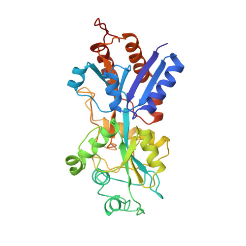
    Electrostatic interactions are among the key forces determining the structure and function of proteins. These are exemplified in the liganded form of the receptor, a phosphate binding protein from Escherichia coli. The phosphate, completely dehydrated and buried in the receptor, is bound by 12 hydrogen bonds as well as a salt link with Arg 135. We have modulated the ionic attraction while preserving the hydrogen bonds by mutating Asp 137, also salt linked to Arg 135, to Asn, Gly or Thr. High-resolution crystallographic analysis revealed that Gly and Thr (but not Asn) mutant proteins have incorporated a more electronegative Cl- in place of the Asp carboxylate. That no dramatic effect on phosphate affinity was produced by these ionic perturbations indicates a major role for hydrogen bonds and other local dipoles in the binding and charge stabilization of ionic ligands.


  • Organizational Affiliation

    Department of Biochemistry, Baylor College of Medicine, Houston, Texas 77030, USA.


Macromolecules
Find similar proteins by:  (by identity cutoff)  |  3D Structure
Entity ID: 1
MoleculeChains Sequence LengthOrganismDetailsImage
PHOSPHATE-BINDING PROTEIN321Escherichia coliMutation(s): 1 
UniProt
Find proteins for P0AG82 (Escherichia coli (strain K12))
Explore P0AG82 
Go to UniProtKB:  P0AG82
Entity Groups  
Sequence Clusters30% Identity50% Identity70% Identity90% Identity95% Identity100% Identity
UniProt GroupP0AG82
Sequence Annotations
Expand
  • Reference Sequence
Experimental Data & Validation

Experimental Data

  • Method: X-RAY DIFFRACTION
  • Resolution: 1.90 Å
  • R-Value Free: 0.218 
  • R-Value Work: 0.190 
  • R-Value Observed: 0.190 
  • Space Group: P 21 21 21
Unit Cell:
Length ( Å )Angle ( ˚ )
a = 41.97α = 90
b = 63.98β = 90
c = 123.97γ = 90
Software Package:
Software NamePurpose
X-PLORmodel building
X-PLORrefinement
SDMSdata reduction
SDMSdata scaling
X-PLORphasing

Structure Validation

View Full Validation Report


Entry History 

Deposition Data

Revision History  (Full details and data files)

  • Version 1.0: 1996-07-11
    Type: Initial release
  • Version 1.1: 2008-03-24
    Changes: Version format compliance
  • Version 1.2: 2011-07-13
    Changes: Version format compliance
  • Version 1.3: 2018-03-21
    Changes: Data collection, Other
  • Version 1.4: 2021-11-03
    Changes: Database references, Derived calculations
  • Version 1.5: 2024-02-14
    Changes: Data collection