1QTI

Acetylcholinesterase (E.C.3.1.1.7)


Experimental Data Snapshot

  • Method: X-RAY DIFFRACTION
  • Resolution: 2.50 Å
  • R-Value Free: 0.236 
  • R-Value Work: 0.192 
  • R-Value Observed: 0.192 

wwPDB Validation   3D Report Full Report


Ligand Structure Quality Assessment 


This is version 1.3 of the entry. See complete history


Literature

Three-dimensional structure of a complex of galanthamine (Nivalin) with acetylcholinesterase from Torpedo californica: implications for the design of new anti-Alzheimer drugs

Bartolucci, C.Perola, E.Pilger, C.Fels, G.Lamba, D.

(2001) Proteins 42: 182-191

  • DOI: https://doi.org/10.1002/1097-0134(20010201)42:2<182::aid-prot50>3.0.co;2-1
  • Primary Citation of Related Structures:  
    1QTI

  • PubMed Abstract: 

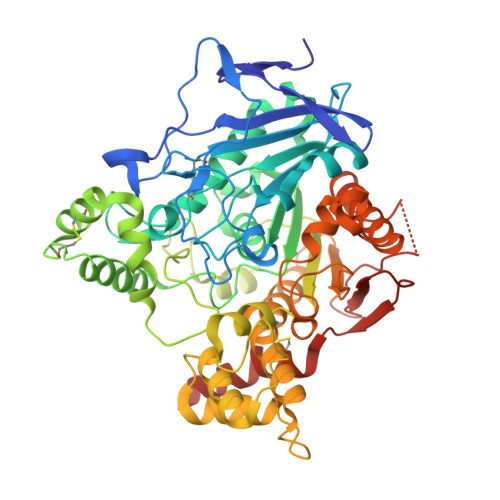
    The 3D structure of a complex of the anti-Alzheimer drug galanthamine with Torpedo californica acetylcholinesterase is reported. Galanthamine, a tertiary alkaloid extracted from several species of Amarylidacae, is so far the only drug that shows a dual activity, being both an acetylcholinesterase inhibitor and an allosteric potentiator of the nicotinic response induced by acetylcholine and competitive agonists. The X-ray structure, at 2.5A resolution, shows an unexpected orientation of the ligand within the active site, as well as unusual protein-ligand interactions. The inhibitor binds at the base of the active site gorge, interacting with both the acyl-binding pocket and the principal quaternary ammonium-binding site. However, the tertiary amine group of galanthamine does not directly interact with Trp84. A docking study using the program AUTODOCK correctly predicts the orientation of galanthamine in the active site. The docked lowest-energy structure has a root mean square deviation of 0.5A with respect to the corresponding crystal structure of the complex. The observed binding mode explains the affinities of a series of structural analogs of galanthamine and provides a rational basis for structure-based drug design of synthetic derivatives with improved pharmacological properties. Proteins 2001;42:182-191.


  • Organizational Affiliation

    Istituto di Strutturistica Chimica G. Giacomello, Monterotondo Stazione, Roma, Italy.


Macromolecules
Find similar proteins by:  (by identity cutoff)  |  3D Structure
Entity ID: 1
MoleculeChains Sequence LengthOrganismDetailsImage
ACETYLCHOLINESTERASE537Tetronarce californicaMutation(s): 0 
EC: 3.1.1.7
UniProt
Find proteins for P04058 (Tetronarce californica)
Explore P04058 
Go to UniProtKB:  P04058
Entity Groups  
Sequence Clusters30% Identity50% Identity70% Identity90% Identity95% Identity100% Identity
UniProt GroupP04058
Sequence Annotations
Expand
  • Reference Sequence
Small Molecules
Ligands 1 Unique
IDChains Name / Formula / InChI Key2D Diagram3D Interactions
GNT
Query on GNT

Download Ideal Coordinates CCD File 
B [auth A](-)-GALANTHAMINE
C17 H21 N O3
ASUTZQLVASHGKV-JDFRZJQESA-N
Binding Affinity Annotations 
IDSourceBinding Affinity
GNT BindingDB:  1QTI Ki: 190 (nM) from 1 assay(s)
IC50: min: 360, max: 1069 (nM) from 6 assay(s)
PDBBind:  1QTI IC50: 360 (nM) from 1 assay(s)
Experimental Data & Validation

Experimental Data

  • Method: X-RAY DIFFRACTION
  • Resolution: 2.50 Å
  • R-Value Free: 0.236 
  • R-Value Work: 0.192 
  • R-Value Observed: 0.192 
  • Space Group: P 31 2 1
Unit Cell:
Length ( Å )Angle ( ˚ )
a = 110.824α = 90
b = 110.824β = 90
c = 136.445γ = 120
Software Package:
Software NamePurpose
DENZOdata reduction
SCALEPACKdata scaling
X-PLORmodel building
X-PLORrefinement
X-PLORphasing

Structure Validation

View Full Validation Report


Ligand Structure Quality Assessment 


Entry History 

Deposition Data

Revision History  (Full details and data files)

  • Version 1.0: 1999-12-29
    Type: Initial release
  • Version 1.1: 2008-04-27
    Changes: Version format compliance
  • Version 1.2: 2011-07-13
    Changes: Version format compliance
  • Version 1.3: 2021-06-02
    Changes: Advisory, Data collection, Derived calculations