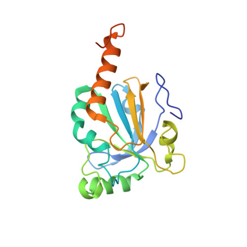
Crystal structure of a multifunctional 2-Cys peroxiredoxin heme-binding protein 23 kDa/proliferation-associated gene product.
Hirotsu, S., Abe, Y., Okada, K., Nagahara, N., Hori, H., Nishino, T., Hakoshima, T.(1999) Proc Natl Acad Sci U S A 96: 12333-12338
- PubMed: 10535922
- DOI: https://doi.org/10.1073/pnas.96.22.12333
- Primary Citation of Related Structures:
1QQ2 - PubMed Abstract:
Heme-binding protein 23 kDa (HBP23), a rat isoform of human proliferation-associated gene product (PAG), is a member of the peroxiredoxin family of peroxidases, having two conserved cysteine residues. Recent biochemical studies have shown that HBP23/PAG is an oxidative stress-induced and proliferation-coupled multifunctional protein that exhibits specific bindings to c-Abl protein tyrosine kinase and heme, as well as a peroxidase activity. A 2.6-A resolution crystal structure of rat HBP23 in oxidized form revealed an unusual dimer structure in which the active residue Cys-52 forms a disulfide bond with conserved Cys-173 from another subunit by C-terminal tail swapping. The active site is largely hydrophobic with partially exposed Cys-173, suggesting a reduction mechanism of oxidized HBP23 by thioredoxin. Thus, the unusual cysteine disulfide bond is involved in peroxidation catalysis by using thioredoxin as the source of reducing equivalents. The structure also provides a clue to possible interaction surfaces for c-Abl and heme. Several significant structural differences have been found from a 1-Cys peroxiredoxin, ORF6, which lacks the C-terminal conserved cysteine corresponding to Cys-173 of HBP23.
Organizational Affiliation:
Department of Molecular Biology, Nara Institute of Science and Technology, 8916-5 Takayama, Ikoma, Nara 630-0101, Japan.