1QOR

CRYSTAL STRUCTURE OF ESCHERICHIA COLI QUINONE OXIDOREDUCTASE COMPLEXED WITH NADPH


Experimental Data Snapshot

  • Method: X-RAY DIFFRACTION
  • Resolution: 2.20 Å
  • R-Value Work: 0.140 
  • R-Value Observed: 0.140 

wwPDB Validation   3D Report Full Report


This is version 1.5 of the entry. See complete history


Literature

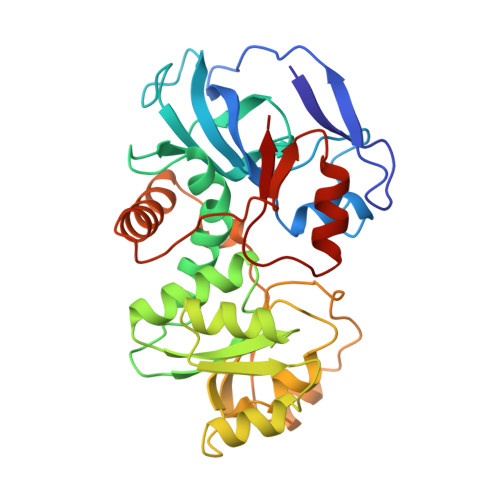
Crystal structure of Escherichia coli QOR quinone oxidoreductase complexed with NADPH.

Thorn, J.M.Barton, J.D.Dixon, N.E.Ollis, D.L.Edwards, K.J.

(1995) J Mol Biol 249: 785-799

  • DOI: https://doi.org/10.1006/jmbi.1995.0337
  • Primary Citation of Related Structures:  
    1QOR

  • PubMed Abstract: 

    The crystal structure of the homodimer of quinone oxidoreductase from Escherichia coli has been determined using the multiple isomorphous replacement method at 2.2 A resolution and refined to an R-factor of 14.1% The crystallographic asymmetric unit contains one functional dimer with the two subunits being related by a non-crystallographic 2-fold symmetry axis. The model consists of two polypeptide chains (residues 2 through 327), one NADPH molecule and one sulphate anion per subunit, and 432 water molecules. Each subunit consists of two domains: a catalytic domain and a nucleotide-binding domain with the NADPH co-factor bound in the cleft between domains. Quinone oxidoreductase has an unusual nucleotide-binding fingerprint motif consisting of the sequence AXXGXXG. The overall structure of quinone oxidoreductase shows strong structural homology to that of horse liver alcohol dehydrogenase.


  • Organizational Affiliation

    Centre for Molecular Structure and Function, Research School of Chemistry, Australian National University, Canberra.


Macromolecules
Find similar proteins by:  (by identity cutoff)  |  3D Structure
Entity ID: 1
MoleculeChains Sequence LengthOrganismDetailsImage
QUINONE OXIDOREDUCTASE
A, B
327Escherichia coliMutation(s): 0 
UniProt
Find proteins for P28304 (Escherichia coli (strain K12))
Explore P28304 
Go to UniProtKB:  P28304
Entity Groups  
Sequence Clusters30% Identity50% Identity70% Identity90% Identity95% Identity100% Identity
UniProt GroupP28304
Sequence Annotations
Expand
  • Reference Sequence
Small Molecules
Ligands 2 Unique
IDChains Name / Formula / InChI Key2D Diagram3D Interactions
NDP
Query on NDP

Download Ideal Coordinates CCD File 
D [auth A],
F [auth B]
NADPH DIHYDRO-NICOTINAMIDE-ADENINE-DINUCLEOTIDE PHOSPHATE
C21 H30 N7 O17 P3
ACFIXJIJDZMPPO-NNYOXOHSSA-N
SO4
Query on SO4

Download Ideal Coordinates CCD File 
C [auth A],
E [auth B]
SULFATE ION
O4 S
QAOWNCQODCNURD-UHFFFAOYSA-L
Experimental Data & Validation

Experimental Data

  • Method: X-RAY DIFFRACTION
  • Resolution: 2.20 Å
  • R-Value Work: 0.140 
  • R-Value Observed: 0.140 
  • Space Group: P 21 21 21
Unit Cell:
Length ( Å )Angle ( ˚ )
a = 107.44α = 90
b = 104.06β = 90
c = 77.45γ = 90
Software Package:
Software NamePurpose
X-PLORmodel building
X-PLORrefinement
X-PLORphasing

Structure Validation

View Full Validation Report


Entry History 

Deposition Data

Revision History  (Full details and data files)

  • Version 1.0: 1995-06-03
    Type: Initial release
  • Version 1.1: 2008-03-03
    Changes: Version format compliance
  • Version 1.2: 2011-07-13
    Changes: Version format compliance
  • Version 1.3: 2013-12-04
    Changes: Non-polymer description
  • Version 1.4: 2017-11-29
    Changes: Derived calculations, Other
  • Version 1.5: 2024-02-14
    Changes: Data collection, Database references, Derived calculations