1QNO

The 3-D structure of a Trichoderma reesei b-mannanase from glycoside hydrolase family 5


Experimental Data Snapshot

  • Method: X-RAY DIFFRACTION
  • Resolution: 2.00 Å
  • R-Value Free: 0.200 
  • R-Value Work: 0.126 

wwPDB Validation   3D Report Full Report


This is version 1.3 of the entry. See complete history


Literature

The Three-Dimensional Structure of a Trichoderma Reesei Beta-Mannanase from Glycoside Hydrolase Family 5.

Sabini, E.Schubert, H.Murshudov, G.Wilson, K.S.Siika-Aho, M.Penttila, M.

(2000) Acta Crystallogr D Biol Crystallogr 56: 3

  • DOI: https://doi.org/10.1107/s0907444999013943
  • Primary Citation of Related Structures:  
    1QNO, 1QNP, 1QNQ, 1QNR, 1QNS

  • PubMed Abstract: 

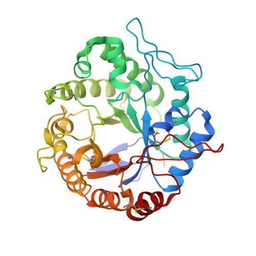
    The crystal structure of the catalytic core domain of beta-mannanase from the fungus Trichoderma reesei has been determined at a resolution of 1.5 A. The structure was solved using the anomalous scattering from a single non-isomorphous platinum complex with two heavy-metal sites in space group P2(1). The map computed with the experimental phases was enhanced by the application of an automated model building and refinement procedure using the amplitudes and experimental phases as observations. This approach is expected to be of more general application. The structure of the native enzyme and complexes with Tris-HCl and mannobiose are also reported: the mannobiose binds in subsites +1 and +2. The structure is briefly compared with that of the homologous beta-mannanase from the bacterium Thermomonospora fusca.


  • Organizational Affiliation

    Structural Biology Laboratory, Department of Chemistry, University of York, Heslington, York YO10 5DD, England.


Macromolecules
Find similar proteins by:  (by identity cutoff)  |  3D Structure
Entity ID: 1
MoleculeChains Sequence LengthOrganismDetailsImage
ENDO-1,4-B-D-MANNANASE344Trichoderma reeseiMutation(s): 0 
EC: 3.2.1.78
UniProt
Find proteins for Q99036 (Hypocrea jecorina (strain ATCC 56765 / BCRC 32924 / NRRL 11460 / Rut C-30))
Explore Q99036 
Go to UniProtKB:  Q99036
Entity Groups  
Sequence Clusters30% Identity50% Identity70% Identity90% Identity95% Identity100% Identity
UniProt GroupQ99036
Sequence Annotations
Expand
  • Reference Sequence
Small Molecules
Experimental Data & Validation

Experimental Data

  • Method: X-RAY DIFFRACTION
  • Resolution: 2.00 Å
  • R-Value Free: 0.200 
  • R-Value Work: 0.126 
  • Space Group: P 1 21 1
Unit Cell:
Length ( Å )Angle ( ˚ )
a = 50.01α = 90
b = 54.27β = 111.31
c = 60.24γ = 90
Software Package:
Software NamePurpose
REFMACrefinement
DENZOdata reduction
SCALEPACKdata scaling

Structure Validation

View Full Validation Report


Entry History 

Deposition Data

Revision History  (Full details and data files)

  • Version 1.0: 2000-10-22
    Type: Initial release
  • Version 1.1: 2013-01-30
    Changes: Database references, Derived calculations, Non-polymer description, Other, Version format compliance
  • Version 1.2: 2019-07-24
    Changes: Data collection, Derived calculations
  • Version 1.3: 2020-07-29
    Type: Remediation
    Reason: Carbohydrate remediation
    Changes: Data collection, Derived calculations, Structure summary