1QKT

MUTANT ESTROGEN NUCLEAR RECEPTOR LIGAND BINDING DOMAIN COMPLEXED WITH ESTRADIOL


Experimental Data Snapshot

  • Method: X-RAY DIFFRACTION
  • Resolution: 2.20 Å
  • R-Value Free: 0.273 
  • R-Value Work: 0.223 
  • R-Value Observed: 0.223 

wwPDB Validation   3D Report Full Report


Ligand Structure Quality Assessment 


This is version 1.2 of the entry. See complete history


Literature

Crystal Structure of a Mutant Heralpha Ligand- Binding Domain Reveals Key Structural Features for the Mechanism of Partial Agonism

Gangloff, M.Ruff, M.Eiler, S.Duclaud, S.Wurtz, J.M.Moras, D.

(2001) J Biol Chem 276: 15059

  • DOI: https://doi.org/10.1074/jbc.M009870200
  • Primary Citation of Related Structures:  
    1QKT, 1QKU

  • PubMed Abstract: 

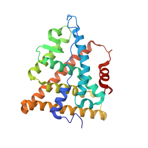
    The crystal structure of a triple cysteine to serine mutant ERalpha ligand-binding domain (LBD), complexed with estradiol, shows that despite the presence of a tightly bound agonist ligand, the protein exhibits an antagonist-like conformation, similar to that observed in raloxifen and 4-hydroxytamoxifen-bound structures. This mutated receptor binds estradiol with wild type affinity and displays transcriptional activity upon estradiol stimulation, but with limited potency (about 50%). This partial activity is efficiently repressed in antagonist competition assays. The comparison with available LBD structures reveals key features governing the positioning of helix H12 and highlights the importance of cysteine residues in promoting an active conformation. Furthermore the present study reveals a hydrogen bond network connecting ligand binding to protein trans conformation. These observations support a dynamic view of H12 positioning, where the control of the equilibrium between two stable locations determines the partial agonist character of a given ligand.


  • Organizational Affiliation

    Institut de Genetique et de Biologie Moleculaire et Cellulaire, Laboratoire de Biologie et de Génomique Structurales 1, rue Laurent Fries, BP 163 67404 Illkirch, France.


Macromolecules
Find similar proteins by:  (by identity cutoff)  |  3D Structure
Entity ID: 1
MoleculeChains Sequence LengthOrganismDetailsImage
ESTRADIOL RECEPTOR248Homo sapiensMutation(s): 3 
UniProt & NIH Common Fund Data Resources
Find proteins for P03372 (Homo sapiens)
Explore P03372 
Go to UniProtKB:  P03372
PHAROS:  P03372
GTEx:  ENSG00000091831 
Entity Groups  
Sequence Clusters30% Identity50% Identity70% Identity90% Identity95% Identity100% Identity
UniProt GroupP03372
Sequence Annotations
Expand
  • Reference Sequence
Small Molecules
Ligands 1 Unique
IDChains Name / Formula / InChI Key2D Diagram3D Interactions
EST
Query on EST

Download Ideal Coordinates CCD File 
B [auth A]ESTRADIOL
C18 H24 O2
VOXZDWNPVJITMN-ZBRFXRBCSA-N
Binding Affinity Annotations 
IDSourceBinding Affinity
EST BindingDB:  1QKT Ki: min: 0.11, max: 100 (nM) from 13 assay(s)
Kd: min: 0.2, max: 100 (nM) from 4 assay(s)
IC50: min: 1.00e-2, max: 46 (nM) from 46 assay(s)
EC50: min: 4.00e-3, max: 10 (nM) from 52 assay(s)
PDBBind:  1QKT Kd: 0.92 (nM) from 1 assay(s)
Binding MOAD:  1QKT Kd: 0.92 (nM) from 1 assay(s)
Experimental Data & Validation

Experimental Data

  • Method: X-RAY DIFFRACTION
  • Resolution: 2.20 Å
  • R-Value Free: 0.273 
  • R-Value Work: 0.223 
  • R-Value Observed: 0.223 
  • Space Group: P 65 2 2
Unit Cell:
Length ( Å )Angle ( ˚ )
a = 58.61α = 90
b = 58.61β = 90
c = 276.02γ = 120
Software Package:
Software NamePurpose
CNSrefinement
DENZOdata reduction
SCALEPACKdata scaling
CNSphasing

Structure Validation

View Full Validation Report


Ligand Structure Quality Assessment 


Entry History 

Deposition Data

Revision History  (Full details and data files)

  • Version 1.0: 2000-08-18
    Type: Initial release
  • Version 1.1: 2011-08-10
    Changes: Atomic model, Database references, Derived calculations, Non-polymer description, Other, Refinement description, Source and taxonomy, Structure summary, Version format compliance
  • Version 1.2: 2022-11-02
    Changes: Database references, Derived calculations, Other