1QHW

PURPLE ACID PHOSPHATASE FROM RAT BONE


Experimental Data Snapshot

  • Method: X-RAY DIFFRACTION
  • Resolution: 2.20 Å
  • R-Value Free: 0.265 
  • R-Value Work: 0.213 
  • R-Value Observed: 0.213 

wwPDB Validation   3D Report Full Report


This is version 2.1 of the entry. See complete history


Literature

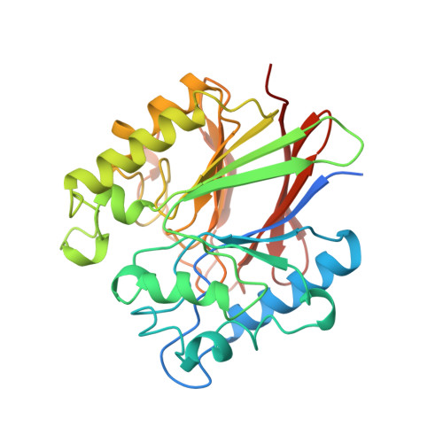
Three-dimensional structure of a mammalian purple acid phosphatase at 2.2 A resolution with a mu-(hydr)oxo bridged di-iron center.

Lindqvist, Y.Johansson, E.Kaija, H.Vihko, P.Schneider, G.

(1999) J Mol Biol 291: 135-147

  • DOI: https://doi.org/10.1006/jmbi.1999.2962
  • Primary Citation of Related Structures:  
    1QHW

  • PubMed Abstract: 

    The crystal structure of purple acid phosphatase from rat bone has been determined by molecular replacement and the structure has been refined to 2.2 A resolution to an R -factor of 21.3 % (R -free 26.5 %). The core of the enzyme consists of two seven-stranded mixed beta-sheets, with each sheet flanked by solvent-exposed alpha-helices on one side. The two sheets pack towards each other forming a beta-sandwich. The di-iron center, located at the bottom of the active-site pocket at one edge of the beta-sandwich, contains a mu-hydroxo or mu-oxo bridge and both metal ions are observed in an almost perfect octahedral coordination geometry. The electron density map indicates that a mu-(hydr)oxo bridge is found in the metal center and that at least one solvent molecule is located in the first coordination sphere of one of the metal ions. The crystallographic study of rat purple acid phosphatase reveals that the mammalian enzymes are very similar in overall structure to the plant enzymes in spite of only 18 % overall sequence identity. In particular, coordination and geometry of the iron cluster is preserved in both enzymes and comparison of the active-sites suggests a common mechanism for the mammalian and plant enzymes. However, significant differences are found in the architecture of the substrate binding pocket.


  • Organizational Affiliation

    Department of Medical Biochemistry & Biophysics, Karolinska Institutet, Stockholm, S-171 77, Sweden. ylva@alfa.mbb.ki.se


Macromolecules
Find similar proteins by:  (by identity cutoff)  |  3D Structure
Entity ID: 1
MoleculeChains Sequence LengthOrganismDetailsImage
PROTEIN (PURPLE ACID PHOSPHATASE)327Rattus norvegicusMutation(s): 0 
Gene Names: ACP5
EC: 3.1.3.2
UniProt
Find proteins for P29288 (Rattus norvegicus)
Explore P29288 
Go to UniProtKB:  P29288
Entity Groups  
Sequence Clusters30% Identity50% Identity70% Identity90% Identity95% Identity100% Identity
UniProt GroupP29288
Sequence Annotations
Expand
  • Reference Sequence
Oligosaccharides

Help

Entity ID: 2
MoleculeChains Length2D Diagram Glycosylation3D Interactions
2-acetamido-2-deoxy-beta-D-glucopyranose-(1-4)-2-acetamido-2-deoxy-beta-D-glucopyranose
B
2N-Glycosylation
Glycosylation Resources
GlyTouCan:  G42666HT
GlyCosmos:  G42666HT
GlyGen:  G42666HT
Experimental Data & Validation

Experimental Data

  • Method: X-RAY DIFFRACTION
  • Resolution: 2.20 Å
  • R-Value Free: 0.265 
  • R-Value Work: 0.213 
  • R-Value Observed: 0.213 
  • Space Group: P 21 21 21
Unit Cell:
Length ( Å )Angle ( ˚ )
a = 69.98α = 90
b = 88.12β = 90
c = 57.07γ = 90
Software Package:
Software NamePurpose
AMoREphasing
CNSrefinement
MOSFLMdata reduction
CCP4data scaling

Structure Validation

View Full Validation Report


Entry History 

Deposition Data

Revision History  (Full details and data files)

  • Version 1.0: 1999-09-15
    Type: Initial release
  • Version 1.1: 2007-10-16
    Changes: Version format compliance
  • Version 1.2: 2011-07-13
    Changes: Non-polymer description, Version format compliance
  • Version 2.0: 2020-07-29
    Type: Remediation
    Reason: Carbohydrate remediation
    Changes: Atomic model, Data collection, Derived calculations, Structure summary
  • Version 2.1: 2023-08-16
    Changes: Data collection, Database references, Refinement description, Structure summary