1QGW

CRYSTAL STRUCTURE OF PHYCOERYTHRIN 545 FROM THE MARINE CRYPTOPHYTE RHODOMONAS CS24


Experimental Data Snapshot

  • Method: X-RAY DIFFRACTION
  • Resolution: 1.63 Å
  • R-Value Free: 0.188 
  • R-Value Work: 0.149 

wwPDB Validation   3D Report Full Report


This is version 1.5 of the entry. See complete history


Literature

Evolution of a light-harvesting protein by addition of new subunits and rearrangement of conserved elements: crystal structure of a cryptophyte phycoerythrin at 1.63-A resolution.

Wilk, K.E.Harrop, S.J.Jankova, L.Edler, D.Keenan, G.Sharples, F.Hiller, R.G.Curmi, P.M.

(1999) Proc Natl Acad Sci U S A 96: 8901-8906

  • DOI: https://doi.org/10.1073/pnas.96.16.8901
  • Primary Citation of Related Structures:  
    1QGW

  • PubMed Abstract: 

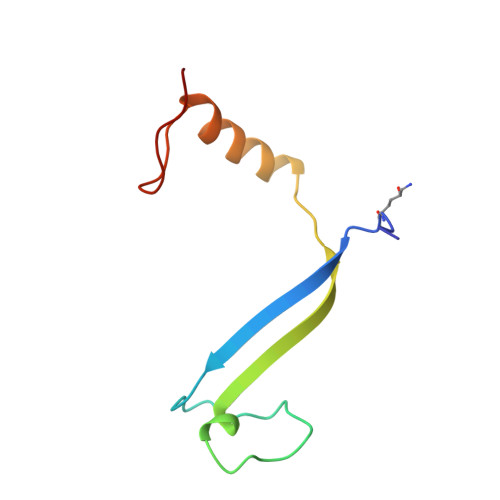
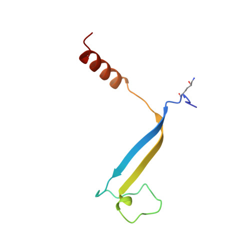
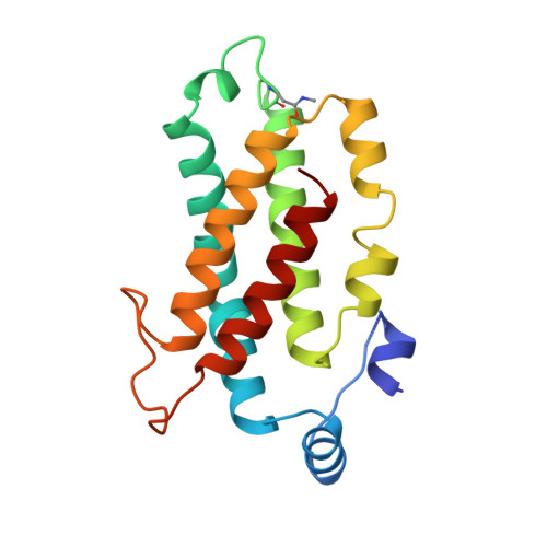
    Cryptophytes are unicellular photosynthetic algae that use a lumenally located light-harvesting system, which is distinct from the phycobilisome structure found in cyanobacteria and red algae. One of the key components of this system is water-soluble phycoerythrin (PE) 545 whose expression is enhanced by low light levels. The crystal structure of the heterodimeric alpha(1)alpha(2)betabeta PE 545 from the marine cryptophyte Rhodomonas CS24 has been determined at 1.63-A resolution. Although the beta-chain structure is similar to the alpha and beta chains of other known phycobiliproteins, the overall structure of PE 545 is novel with the alpha chains forming a simple extended fold with an antiparallel beta-ribbon followed by an alpha-helix. The two doubly linked beta50/beta61 chromophores (one on each beta subunit) are in van der Waals contact, suggesting that exciton-coupling mechanisms may alter their spectral properties. Each alpha subunit carries a covalently linked 15,16-dihydrobiliverdin chromophore that is likely to be the final energy acceptor. The architecture of the heterodimer suggests that PE 545 may dock to an acceptor protein via a deep cleft and that energy may be transferred via this intermediary protein to the reaction center.


  • Organizational Affiliation

    Initiative in Biomolecular Structure, School of Physics, University of New South Wales, Sydney, NSW 2052, Australia.


Macromolecules
Find similar proteins by:  (by identity cutoff)  |  3D Structure
Entity ID: 1
MoleculeChains Sequence LengthOrganismDetailsImage
PROTEIN (CRYPTOPHYTAN PHYCOERYTHRIN (ALPHA-1 CHAIN))76Rhodomonas sp. CS24Mutation(s): 1 
UniProt
Find proteins for Q00433 (Rhodomonas sp. (strain CS 24))
Explore Q00433 
Go to UniProtKB:  Q00433
Entity Groups  
Sequence Clusters30% Identity50% Identity70% Identity90% Identity95% Identity100% Identity
UniProt GroupQ00433
Sequence Annotations
Expand
  • Reference Sequence
Find similar proteins by:  (by identity cutoff)  |  3D Structure
Entity ID: 2
MoleculeChains Sequence LengthOrganismDetailsImage
PROTEIN (CRYPTOPHYTAN PHYCOERYTHRIN (ALPHA-2 CHAIN))67Rhodomonas sp. CS24Mutation(s): 1 
UniProt
Find proteins for P30943 (Rhodomonas sp. (strain CS 24))
Explore P30943 
Go to UniProtKB:  P30943
Entity Groups  
Sequence Clusters30% Identity50% Identity70% Identity90% Identity95% Identity100% Identity
UniProt GroupP30943
Sequence Annotations
Expand
  • Reference Sequence
Find similar proteins by:  (by identity cutoff)  |  3D Structure
Entity ID: 3
MoleculeChains Sequence LengthOrganismDetailsImage
PROTEIN (CRYPTOPHYTAN PHYCOERYTHRIN (BETA CHAIN))
C, D
177Rhodomonas sp. CS24Mutation(s): 1 
UniProt
Find proteins for P27198 (Rhodomonas sp. (strain CS 24))
Explore P27198 
Go to UniProtKB:  P27198
Entity Groups  
Sequence Clusters30% Identity50% Identity70% Identity90% Identity95% Identity100% Identity
UniProt GroupP27198
Sequence Annotations
Expand
  • Reference Sequence
Small Molecules
Ligands 4 Unique
IDChains Name / Formula / InChI Key2D Diagram3D Interactions
PEB
Query on PEB

Download Ideal Coordinates CCD File 
I [auth C]
J [auth C]
K [auth C]
M [auth D]
N [auth D]
I [auth C],
J [auth C],
K [auth C],
M [auth D],
N [auth D],
O [auth D]
PHYCOERYTHROBILIN
C33 H40 N4 O6
NKCBCVIFPXGHAV-WAVSMFBNSA-N
DBV
Query on DBV

Download Ideal Coordinates CCD File 
F [auth A],
G [auth B]
15,16-DIHYDROBILIVERDIN
C33 H36 N4 O6
ZQHDSLZHMAUUQK-ZTYGKHTCSA-N
CL
Query on CL

Download Ideal Coordinates CCD File 
H [auth C]CHLORIDE ION
Cl
VEXZGXHMUGYJMC-UHFFFAOYSA-M
MG
Query on MG

Download Ideal Coordinates CCD File 
E [auth A],
L [auth D]
MAGNESIUM ION
Mg
JLVVSXFLKOJNIY-UHFFFAOYSA-N
Modified Residues  2 Unique
IDChains TypeFormula2D DiagramParent
LYZ
Query on LYZ
A
L-PEPTIDE LINKINGC6 H14 N2 O3LYS
MEN
Query on MEN
C, D
L-PEPTIDE LINKINGC5 H10 N2 O3ASN
Experimental Data & Validation

Experimental Data

  • Method: X-RAY DIFFRACTION
  • Resolution: 1.63 Å
  • R-Value Free: 0.188 
  • R-Value Work: 0.149 
  • Space Group: P 21 21 21
Unit Cell:
Length ( Å )Angle ( ˚ )
a = 63.03α = 90
b = 82.63β = 90
c = 89.55γ = 90
Software Package:
Software NamePurpose
AMoREphasing
ARPmodel building
REFMACrefinement
DENZOdata reduction
SCALEPACKdata scaling
ARP/wARPmodel building

Structure Validation

View Full Validation Report


Entry History 

Deposition Data

Revision History  (Full details and data files)

  • Version 1.0: 1999-05-19
    Type: Initial release
  • Version 1.1: 2008-04-26
    Changes: Version format compliance
  • Version 1.2: 2011-07-13
    Changes: Version format compliance
  • Version 1.3: 2018-03-07
    Changes: Advisory, Data collection
  • Version 1.4: 2022-12-21
    Changes: Advisory, Database references, Derived calculations
  • Version 1.5: 2023-09-20
    Changes: Data collection, Refinement description