Kinetic and structural characteristics of the inhibition of enoyl (acyl carrier protein) reductase by triclosan.
Ward, W.H., Holdgate, G.A., Rowsell, S., McLean, E.G., Pauptit, R.A., Clayton, E., Nichols, W.W., Colls, J.G., Minshull, C.A., Jude, D.A., Mistry, A., Timms, D., Camble, R., Hales, N.J., Britton, C.J., Taylor, I.W.(1999) Biochemistry 38: 12514-12525
- PubMed: 10493822
- DOI: https://doi.org/10.1021/bi9907779
- Primary Citation of Related Structures:
1QG6 - PubMed Abstract:
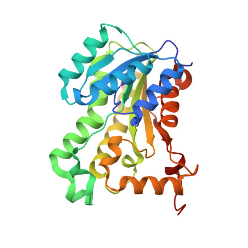
Triclosan is used widely as an antibacterial agent in dermatological products, mouthwashes, and toothpastes. Recent studies imply that antibacterial activity results from binding to enoyl (acyl carrier protein) reductase (EACPR, EC 1.3.1.9). We first recognized the ability of triclosan to inhibit EACPR from Escherichia coli in a high throughput screen where the enzyme and test compound were preincubated with NAD(+), which is a product of the reaction. The concentration of triclosan required for 50% inhibition approximates to 50% of the enzyme concentration, indicating that the free compound is depleted by binding to EACPR. With no preincubation or added NAD(+), the degree of inhibition by 150 nM triclosan increases gradually over several minutes. The onset of inhibition is more rapid when NAD(+) is added. Gel filtration and mass spectrometry show that inhibition by triclosan is reversible. Steady-state assays were designed to avoid depletion of free inhibitor and changes in the degree of inhibition. The results suggest that triclosan binds to E-NAD(+) complex, with a dissociation constant around 20-40 pM. Triclosan follows competitive kinetics with respect to NADH, giving an inhibition constant of 38 pM at zero NADH and saturating NAD(+). Uncompetitive kinetics are observed when NAD(+) is varied, giving an inhibition constant of 22 pM at saturating NAD(+). By following regain of catalytic activity after dilution of EACPR that had been preincubated with triclosan and NAD(+), the rate constant for dissociation of the inhibitor (k(off)) is measured as 1.9 x 10(-4) s(-1). The association rate constant (k(on)) is estimated as 2.6 x 10(7) s(-1) M(-1) by monitoring the onset of inhibition during assays started by addition of EACPR. As expected, the ratio k(off)/k(on) = 7.1 pM is similar to the inhibition constants from the steady-state studies. The crystal structure of E. coli EACPR in a complex with coenzyme and triclosan has been determined at 1.9 A resolution, showing that this compound binds in a similar site to the diazaborine inhibitors. The high affinity of triclosan appears to be due to structural similarity to a tightly bound intermediate in catalysis.
Organizational Affiliation:
AstraZeneca, Mereside, Macclesfield, Cheshire, U.K. walter.ward@astrazeneca.com