1QFX

PH 2.5 ACID PHOSPHATASE FROM ASPERGILLUS NIGER


Experimental Data Snapshot

  • Method: X-RAY DIFFRACTION
  • Resolution: 2.40 Å
  • R-Value Free: 0.225 
  • R-Value Work: 0.183 

wwPDB Validation   3D Report Full Report


Ligand Structure Quality Assessment 


This is version 2.1 of the entry. See complete history


Literature

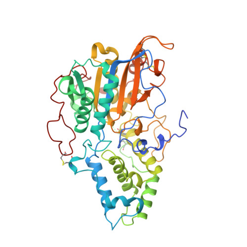
Crystal structure of Aspergillus niger pH 2.5 acid phosphatase at 2. 4 A resolution.

Kostrewa, D.Wyss, M.D'Arcy, A.van Loon, A.P.

(1999) J Mol Biol 288: 965-974

  • DOI: https://doi.org/10.1006/jmbi.1999.2736
  • Primary Citation of Related Structures:  
    1QFX

  • PubMed Abstract: 

    The crystal structure of Aspergillus niger pH 2.5 acid phosphatase (EC 3.1.3.2) has been determined at 2.4 A resolution. In the crystal, two dimers form a tetramer in which the active sites are easily accessible to substrates. The main contacts in the dimer come from the N termini, each lying on the surface of the neighbouring molecule. The monomer consists of two domains, with the active site located at their interface. The active site has a highly conserved catalytic center and a charge distribution, which explains the highly acidic pH optimum and the broad substrate specificity of the enzyme.


  • Organizational Affiliation

    F. Hoffmann-La Roche Ltd, B/65/R312, Basel, 4070, Switzerland. dirk.kostrewa@roche.com


Macromolecules
Find similar proteins by:  (by identity cutoff)  |  3D Structure
Entity ID: 1
MoleculeChains Sequence LengthOrganismDetailsImage
PROTEIN (PH 2.5 ACID PHOSPHATASE)
A, B
460Aspergillus nigerMutation(s): 0 
EC: 3.1.3.8
UniProt
Find proteins for P34755 (Aspergillus awamori)
Explore P34755 
Go to UniProtKB:  P34755
Entity Groups  
Sequence Clusters30% Identity50% Identity70% Identity90% Identity95% Identity100% Identity
UniProt GroupP34755
Sequence Annotations
Expand
  • Reference Sequence
Oligosaccharides

Help

Entity ID: 2
MoleculeChains Length2D Diagram Glycosylation3D Interactions
alpha-D-mannopyranose-(1-2)-alpha-D-mannopyranose-(1-2)-alpha-D-mannopyranose-(1-3)-[alpha-D-mannopyranose-(1-6)]beta-D-mannopyranose-(1-4)-2-acetamido-2-deoxy-beta-D-glucopyranose-(1-4)-2-acetamido-2-deoxy-beta-D-glucopyranose
C
7N-Glycosylation
Glycosylation Resources
GlyTouCan:  G46836GH
GlyCosmos:  G46836GH
GlyGen:  G46836GH
Entity ID: 3
MoleculeChains Length2D Diagram Glycosylation3D Interactions
alpha-D-mannopyranose-(1-3)-[alpha-D-mannopyranose-(1-6)]beta-D-mannopyranose-(1-4)-2-acetamido-2-deoxy-beta-D-glucopyranose-(1-4)-2-acetamido-2-deoxy-beta-D-glucopyranose
D
5N-Glycosylation
Glycosylation Resources
GlyTouCan:  G22768VO
GlyCosmos:  G22768VO
GlyGen:  G22768VO
Small Molecules
Ligands 3 Unique
IDChains Name / Formula / InChI Key2D Diagram3D Interactions
NAG
Query on NAG

Download Ideal Coordinates CCD File 
E [auth A],
F [auth A],
O [auth B],
P [auth B]
2-acetamido-2-deoxy-beta-D-glucopyranose
C8 H15 N O6
OVRNDRQMDRJTHS-FMDGEEDCSA-N
SO4
Query on SO4

Download Ideal Coordinates CCD File 
G [auth A]
H [auth A]
I [auth A]
J [auth A]
K [auth A]
G [auth A],
H [auth A],
I [auth A],
J [auth A],
K [auth A],
Q [auth B],
R [auth B],
S [auth B],
T [auth B],
U [auth B],
V [auth B]
SULFATE ION
O4 S
QAOWNCQODCNURD-UHFFFAOYSA-L
GOL
Query on GOL

Download Ideal Coordinates CCD File 
L [auth A]
M [auth A]
N [auth A]
W [auth B]
X [auth B]
L [auth A],
M [auth A],
N [auth A],
W [auth B],
X [auth B],
Y [auth B]
GLYCEROL
C3 H8 O3
PEDCQBHIVMGVHV-UHFFFAOYSA-N
Experimental Data & Validation

Experimental Data

  • Method: X-RAY DIFFRACTION
  • Resolution: 2.40 Å
  • R-Value Free: 0.225 
  • R-Value Work: 0.183 
  • Space Group: P 61 2 2
Unit Cell:
Length ( Å )Angle ( ˚ )
a = 170.6α = 90
b = 170.6β = 90
c = 201.15γ = 120
Software Package:
Software NamePurpose
XDSdata scaling
XDSdata reduction
SHELXSphasing
SHARPphasing
CCP4model building
X-PLORrefinement
CCP4phasing

Structure Validation

View Full Validation Report


Ligand Structure Quality Assessment 


Entry History 

Deposition Data

Revision History  (Full details and data files)

  • Version 1.0: 2000-04-19
    Type: Initial release
  • Version 1.1: 2008-04-26
    Changes: Version format compliance
  • Version 1.2: 2011-07-13
    Changes: Derived calculations, Version format compliance
  • Version 2.0: 2020-07-29
    Type: Remediation
    Reason: Carbohydrate remediation
    Changes: Atomic model, Data collection, Derived calculations, Structure summary
  • Version 2.1: 2023-12-27
    Changes: Data collection, Database references, Structure summary