1QD3

HIV-1 TAR RNA/NEOMYCIN B COMPLEX


Experimental Data Snapshot

  • Method: SOLUTION NMR
  • Conformers Calculated: 100 
  • Conformers Submitted: 17 
  • Selection Criteria: LOWEST ENERGY, AGREEMENT WITH EXPERIMENTAL DATA 

wwPDB Validation   3D Report Full Report


This is version 2.1 of the entry. See complete history


Literature

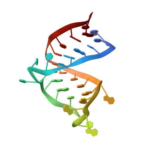
Structural rearrangements of HIV-1 Tat-responsive RNA upon binding of neomycin B.

Faber, C.Sticht, H.Schweimer, K.Rosch, P.

(2000) J Biol Chem 275: 20660-20666

  • DOI: https://doi.org/10.1074/jbc.M000920200
  • Primary Citation of Related Structures:  
    1QD3

  • PubMed Abstract: 

    Binding of human immunodeficiency virus type 1 (HIV-1) transactivator (Tat) protein to Tat-responsive RNA (TAR) is essential for viral replication and is considered a promising starting point for the design of anti-HIV drugs. NMR spectroscopy indicated that the aminoglycosides neomycin B and ribostamycin bind to TAR and that neomycin is able to inhibit Tat binding to TAR. The solution structure of the neomycin-bound TAR has been determined by NMR spectroscopy. Chemical shift mapping and intermolecular nuclear Overhauser effects define the binding region of the aminoglycosides on TAR and give strong evidence for minor groove binding. Based on 15 nuclear Overhauser effect-derived intermolecular distance restraints, a model structure of the TAR-neomycin complex was calculated. Neomycin is bound in a binding pocket formed by the minor groove of the lower stem and the uridine-rich bulge of TAR, which adopts a conformation different from those known. The neamine core of the aminoglycoside (rings I and II) is covered with the bulge, explaining the inhibition of Tat by an allosteric mechanism. Neomycin reduces the volume of the major groove in which Tat is bound and thus impedes essential protein-RNA contacts.


  • Organizational Affiliation

    Lehrstuhl für Biopolymere, Universität Bayreuth, D-95447 Bayreuth, Germany.


Macromolecules
Find similar nucleic acids by:  (by identity cutoff)  |  3D Structure
Entity ID: 1
MoleculeChains LengthOrganismImage
HIV-1 TAR RNA29Human immunodeficiency virus 1
Sequence Annotations
Expand
  • Reference Sequence
Oligosaccharides

Help

Entity ID: 2
MoleculeChains Length2D Diagram Glycosylation3D Interactions
2,6-diamino-2,6-dideoxy-beta-L-idopyranose-(1-3)-alpha-D-ribofuranose
B
2N/A
Glycosylation Resources
GlyTouCan:  G90578XL
GlyCosmos:  G90578XL
Small Molecules
Ligands 2 Unique
IDChains Name / Formula / InChI Key2D Diagram3D Interactions
BDG
Query on BDG

Download Ideal Coordinates CCD File 
C [auth A]2,6-diamino-2,6-dideoxy-alpha-D-glucopyranose
C6 H14 N2 O4
SQTHUUHOUPJYLK-UKFBFLRUSA-N
CYY
Query on CYY

Download Ideal Coordinates CCD File 
D [auth A]2-DEOXYSTREPTAMINE
C6 H14 N2 O3
DTFAJAKTSMLKAT-JDCCYXBGSA-N
Binding Affinity Annotations 
IDSourceBinding Affinity
RIB PDBBind:  1QD3 Kd: 5900 (nM) from 1 assay(s)
Experimental Data & Validation

Experimental Data

  • Method: SOLUTION NMR
  • Conformers Calculated: 100 
  • Conformers Submitted: 17 
  • Selection Criteria: LOWEST ENERGY, AGREEMENT WITH EXPERIMENTAL DATA 

Structure Validation

View Full Validation Report


Entry History 

Deposition Data

Revision History  (Full details and data files)

  • Version 1.0: 2000-07-12
    Type: Initial release
  • Version 1.1: 2008-04-26
    Changes: Version format compliance
  • Version 1.2: 2011-07-13
    Changes: Version format compliance
  • Version 2.0: 2020-07-29
    Type: Remediation
    Reason: Carbohydrate remediation
    Changes: Advisory, Atomic model, Data collection, Derived calculations, Structure summary
  • Version 2.1: 2023-12-27
    Changes: Data collection, Database references, Derived calculations, Structure summary