Kinetic and crystallographic studies on the active site Arg289Lys mutant of flavocytochrome b2 (yeast L-lactate dehydrogenase)
Mowat, C.G., Beaudoin, I., Durley, R.C.E., Barton, J.D., Pike, A.D., Chen, Z.-W., Reid, G.A., Chapman, S.K., Mathews, F.S., Lederer, F.(2000) Biochemistry 39: 3266-3275
- PubMed: 10727218
- DOI: https://doi.org/10.1021/bi9925975
- Primary Citation of Related Structures:
1QCW - PubMed Abstract:
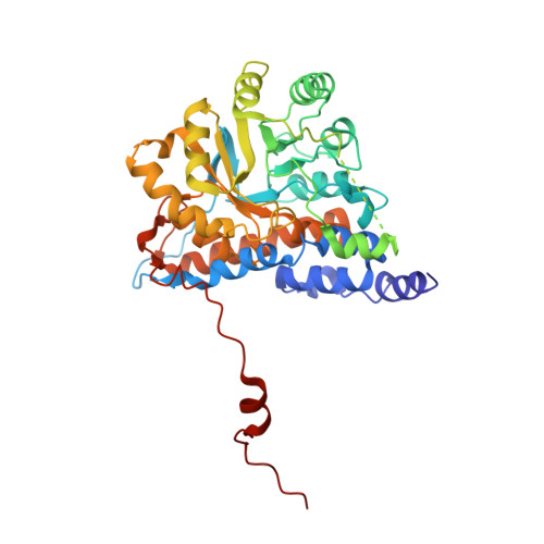
Flavocytochrome b(2) from Saccharomyces cerevisiae couples L-lactate dehydrogenation to cytochrome c reduction. The crystal structure of the native yeast enzyme has been determined [Xia, Z.-X., and Mathews, F. S. (1990) J. Mol. Biol. 212, 837-863] as well as that of the sulfite adduct of the recombinant enzyme produced in Escherichia coli [Tegoni, M., and Cambillau, C. (1994) Protein Sci. 3, 303-313]; several key active site residues were identified. In the sulfite adduct crystal structure, Arg289 adopts two alternative conformations. In one of them, its side chain is stacked against that of Arg376, which interacts with the substrate; in the second orientation, the R289 side chain points toward the active site. This residue has now been mutated to lysine and the mutant enzyme, R289K-b(2), characterized kinetically. Under steady-state conditions, kinetic parameters (including the deuterium kinetic isotope effect) indicate the mutation affects k(cat) by a factor of about 10 and k(cat)/K(M) by up to nearly 10(2). Pre-steady-state kinetic analysis of flavin and heme reduction by lactate demonstrates that the latter is entirely limited by flavin reduction. Inhibition studies on R289K-b(2) with a range of compounds show a general rise in K(i) values relative to that of wild-type enzyme, in line with the elevation of the K(M) for L-lactate in R289K-b(2); they also show a change in the pattern of inhibition by pyruvate and oxalate, as well as a loss of the inhibition by excess substrate. Altogether, the kinetic studies indicate that the mutation has altered the first step of the catalytic cycle, namely, flavin reduction; they suggest that R289 plays a role both in Michaelis complex and transition-state stabilization, as well as in ligand binding to the active site when the flavin is in the semiquinone state. In addition, it appears that the mutation has not affected electron transfer from fully reduced flavin to heme, but may have slowed the second intramolecular ET step, namely, transfer from flavin semiquinone to heme b(2). Finally, the X-ray crystal structure of R289K-b(2), with sulfite bound at the active site, has been determined to 2.75 A resolution. The lysine side chain at position 289 is well-defined and in an orientation that corresponds approximately to one of the alternative conformations observed in the structure of the recombinant enzyme-sulfite complex [Tegoni, M., and Cambillau, C. (1994) Protein Sci. 3, 303-313]. Comparisons between the R289K-b(2) and wild-type structures allow the kinetic results to be interpreted in a structural context.
Organizational Affiliation:
Department of Chemistry, University of Edinburgh, West Mains Road, Edinburgh EH9 3JJ, Scotland, U.K.